Meiosis and Fertilization
Learning Objectives
After completing the lab, the student will be able to:
- Interpret a karyotype;
- Determine karyotype abnormalities and identify an associated disorder or syndrome.
Introduction
In order for a sexually reproducing organism to create offspring that have the proper number of chromosomes for their species, the sperm and egg have to have half the usual number of chromosomes (if sperm and eggs had the full number, each offspring would end up with twice the proper number after they merged). The process that creates these specialized cells (gametes) is called meiosis. Like regular mitosis, it starts with the number of chromosomes in a standard cell being doubled, and then dividing to reduce them back to the starting number. However, it then divides again to create gametes with have that number.
A sperm or egg is called haploid because it has half the normal number of chromosomes (one copy of each chromosome, not a pair of each). Normal cells that have pairs of each type are called diploid.
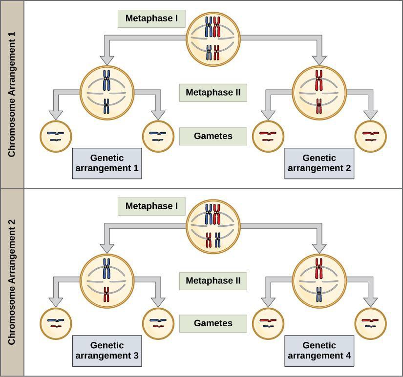
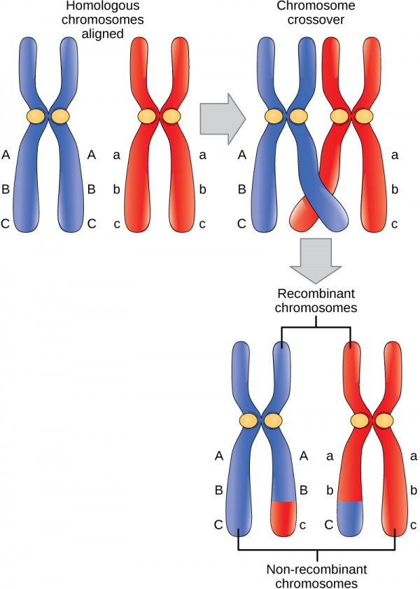
Another key function of meiosis is to mix up the genes that are passed on to each offspring, so that no two offspring get the same (unless they are identical twins). This is accomplished by two steps, one of which randomizes which member of each pair of chromosomes possessed by the parent, gets sent to a gamete that is being created. The reproducing organism has genes that originated in both its mother and father; when this step of re-assortment happens, those that originated from its mother and father don’t stay together, but randomly mix with each other. The other step is called crossing over, and involves intertwining of pieces of chromosomes from the mother and father, so that they exchange chromosomal material. The two chromosomes may swap tips, etc. While re-assortment of chromosomes enabled genes from the mom and dad to be mixed, crossover allows genes on the same chromosome to be separated from each other in the formation of gametes. In general, having a large variety of different mixings of genes within a species, is considered beneficial. This is because it gives different combinations of traits that a species can use to respond to changes in its environment.
An intending parent producing sperm or eggs, has chromosomes from both its mother and father that it will pass into those gametes, and which will thence pass into its own young when it reproduces. The versions of the same chromosome from the parent’s mother and father are called homologous chromosomes. When they get copied in preparation for the double division of the cell in meiosis, the copies and originals are called sister chromatids.
This video demonstrates the sperm producing cells of a male crane fly, a species with only 8 chromosomes per cell during meiotic division.
There are several malfunctions that can occur during meiosis. Nondisjunction occurs when the sister chromatids in tetrads (groups of four) separate unevenly. Nondisjunction results in one cell getting an extra chromosome while another cell is missing a chromosome. Nondisjunction is associated with certain human genetic disorders. For example, Down syndrome is usually caused by nondisjunction that results in three copies of chromosome 21. Translocations can occur when chromosomes exchange genetic information with nonhomologous chromosomes. For a more detailed list, see this web page: https://www.genome.gov/11508982/chromosome-abnormalities-fact-sheet/
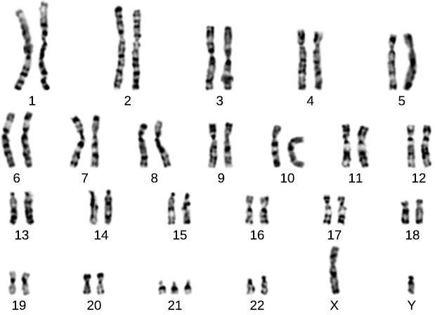
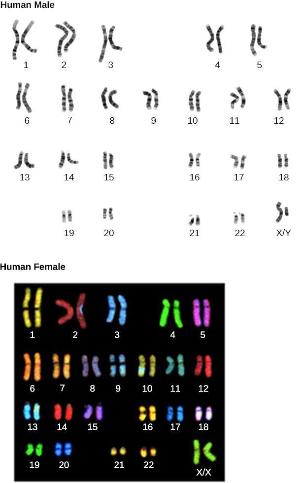
Karyotypes (chromosome spreads) are made by stopping cells in mitosis with a chemical and then staining. A picture is taken through a microscope and then digitally enlarged to see the chromosomal banding, or G-bands. Dark and light banding patterns help identify chromosomes and alterations to normal chromosomes. The chromosome spreads can be seen in Figure 13.7. Human females have two X chromosomes, and males have one X and one Y.
Safety Precautions
None
Materials
- Images of karyotypes
For this activity, you will work in pairs.
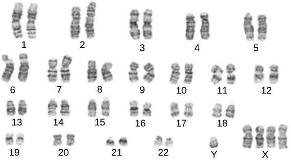
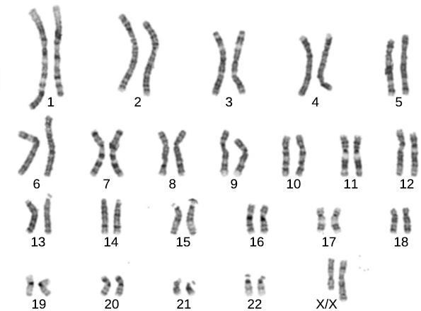
Procedure
Step 1: Examine the three karyotypes 1, 2, and 3 shown in Figure 13.8, Figure 13.9, and Figure 13.10 respectively (below). Compare these karyotypes to the normal karyotypes shown in Figure 13.7 (above). Can you tell if the individual is female, male, or indeterminate (does not have a normal distribution of sex chromosomes)? Record any abnormalities in your notebook, and research the meaning of the changes in chromosome number or appearance.