Lab 5A: Viewing of Prokaryotic Cells by Microscope
Learning Objectives
After completing the lab, the student will be able to:
- Make wet mounts of bacteria, plant and animal cells and view them under the microscope;
- Observe and identify differences between cells and cell structures under low and high magnification and record your observations.
Lab 5A: Viewing of prokaryotic cells by microscope
Introduction
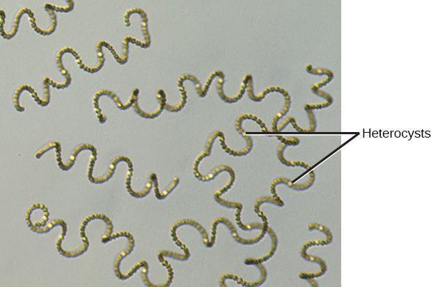
Prokaryotes, unicellular organisms lacking a nucleus, include cyanobacteria (formerly blue-green algae. This name is now considered inaccurate because algae are eukaryotes). Cyanobacteria, like those shown in Figure 4.1, are photoautotrophs—organisms that carry out photosynthesis by using light energy, water, and carbon dioxide from the air and converting to sugars, and providing oxygen to the atmosphere as a waste product. Cyanobacteria contain pigments capable of capturing light energy but do not contain chloroplasts, the organelles used by plants to perform photosynthesis. Cyanobacteria are single-celled organisms, but some can form colonies that may include cells arranged in a long strand or filament.
Safety Precautions
- Be careful when handling glass slides; the edges may be sharp.
- Observe proper use of the microscope; avoid handling the electric cord with wet hands.
- Do not use the coarse adjustment knob of the microscope when using the 40x objective
- Inform your teacher immediately of any broken glassware, as it could cause injuries.
- Wash your hands with soap and water after handling live organisms.
For this activity, you will work in pairs.
Materials
- Light compound microscope
- Lens paper
- Prepared slide of Anabaena
- Special slide with stage micrometer
Procedure
Step 1: View the prepared slide of the cyanobacterium Anabaena. They will be scattered about the image with lots of space in between. Find the largest specimen of Anabaena you can, and center it in the slide (not at the tip of the pointer, at the center of field of view)
Step 2: Draw what you see in your lab notebook, using the 4x, 10x, and 40x objective lenses. Draw the whole view inside a bounding circle, using coarse details, but provide fine details for the central 10% or so of the image.
Step 3: Describe the appearance (shape, color, etc) of an Anabaena filament. Estimate the total number of cells that are visible in that filament.
