Introduction of Identification of Stages of Mitosis
Learning Objectives
After completing the lab, the student will be able to:
- Recognize the stages of mitosis in cells.
- Find cells of each type in a microscope slide.
Introduction
A fundamental property of cells of multicellular organisms is mitosis, the creation of new cells by splitting of existing ones. You may recall from Cell Theory that all cells arise from the splitting of previously existing ones. A simple example in humans is our continuous shedding of skin cells and their replacement by new skin cells. Mitosis is also vital for growth and development of embryos. Many single-celled organisms also perform mitosis- which is their means of reproduction.
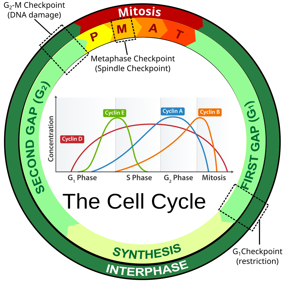
Mitosis is only a small part of the overall life cycle of a cell (See Figure 13.1), called the cell cycle. The bulk of the cell cycle is spent in a period known as interphase, in which the cell is not actively splitting. Interphase is further broken down in to 3 distinct phases: G1 (Gap 1), S (Synthesis) and G2 (Gap 2). G1 is the phase of growth when the cell is accumulating resources to live and grow. After attaining a certain size and having amassed enough raw materials, a checkpoint is reached where the cell uses biochemical markers to decide if it should proceed to S phase. During S phase, the cell copies the DNA in the nucleus in preparation for the cell eventually dividing. At the end of S phase, each chromosome consists of two identical sister chromatids (copies) joined at the centromere. When the DNA synthesis is complete, the cell continues on to the second growth phase called G2. Another checkpoint takes place at the end of G2 to ensure the fidelity of the replicated DNA and to re-establish the success of the cell’s capacity to divide in the environment. If conditions are favorable, the cell continues on to splitting (mitosis). Other structures, such as organelles, are replicated during the G1 portion of the cell cycle (See Figure 13.1).
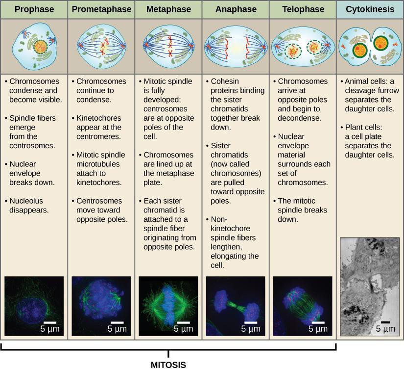
Mitosis is subdivided into five phases. During prophase, the chromosomes coil up, and sister chromatids become visible under a microscope. The nuclear membrane surrounding the chromosomes also disappears. In metaphase, the sister chromatids align in the center of the cell, attached to spindle fibers. During anaphase, the sister chromatids separate and move to opposite poles of the cell. In telophase, the chromosomes arrive at the poles and begin to decondense while the nucleus reforms. Figure 13.3 makes it look like the phases are very distinct. The phases, however, transition without stopping.
Safety Precautions
- Be careful handling glass slides, as the edges may be sharp.
- Observe proper use of the microscope; avoid handling the electric cord with wet hands.
- Do not use the coarse adjustment knob of the microscope at higher magnifications.
- There is a separate marked disposal for sharp objects like broken glass. If you cannot locate it, inform your Instructor/Lab Technician immediately of any broken glassware, as it could cause serious injuries.
Materials
- Prepared slide of onion root tip cells
- Video demonstrating identification of stages of mitosis in Whitefish (early in embryonic development).
For this activity, you will work in pairs.
Procedure
Step 1: Examine slides in a microscope set up by the instructor. View and describe in your lab notebook, distinguishing marks for interphase, prophase, metaphase, anaphase, and telophase phases of mitosis. Do the same for cells in cytokinesis.
Step 2: Identify phases the instructor points to in the slides in the microscope.
