Lab 2c Depth of Field and Wet Mount of Specimens
Learning Objectives
After completing the lab, the student will be able to:
- Explain or describe the depth of field;
- Construct a temporary wet mount , when given a slide, coverslip, and specimen.
Introduction
Preparation of microscope slides varies in complexity, sometimes requiring stains to view items that are difficult to see. However, many simple everyday materials are easy to view without much preparation.
Safety Precautions
- Handle glass slides with care.
- Dispose of specimens as necessary.
- Dispose of coverslips in a broken-glass container.
Methods
- Compound microscope
- Slides with crossed colored fibers or crossed strands of hair
- Clean slides
- Coverslips
- Elodea, onion skin, pond water, or other samples
For this activity, you will work in pairs.
Procedure 1
As you work with a microscope, you will notice that even when an object is in focus, you can continue to adjust the focus and see new things in the lenses. This is because most specimens are very thick. As you move up and down with the stage, different planes of the object come into focus. The depth of field refers to how thick each plane is. It tells you how much of the full depth range of a specimen is in focus at any given time. Although you may think that a higher depth of field is more useful because it allows you to see more objects, most scientists try to make the depth of field as narrow as possible. This prevents features of the image from other focal planes from interfering with the observations.
Step 1: Maneuver the slide of colored threads under scanning power (4x objective) so the cross-point of the threads is center.
Step 2: Switch to 10x. Using the fine focus knob, determine which color of threat is sandwiched between the other two. Record in your lab notebook.
-
-
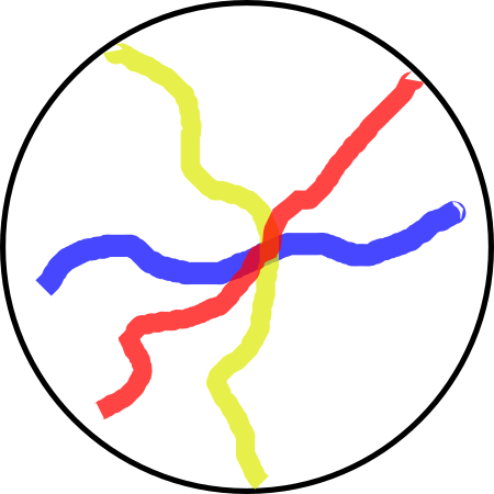
Figure 2.5: Colored threads with cross-points at the center of the field of view. Licensed CC-BY-NC-SA by Jeremy Seto in Biology OER2Procedure 1
-
Procedure 2
The specimens you use on your microscope can either be wet-mounted or dry-mounted. A wet-mount refers to living tissues that are placed on a slide with an aqueous solution to keep them wet. Usually, a coverslip is placed on top of the specimen to flatten the specimen onto the slide. A dry-mount refers to preserved tissue that has been fixed and stained on a slide. This technique allows you to preserve specimens for a long time, and it also allows you to add chemicals to increase the contrast of a specimen from its background.
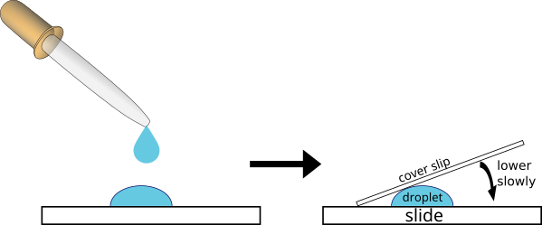
Step 1: You will now observe samples of living organisms provided by your teacher. If using a sample that is not in water, add a drop of water to the specimen. Place a cover slip at an angle so that one edge touches the drop (Fig 2.6). Slowly lower the coverslip onto the specimen. Avoid trapping air bubbles under the coverslip.
Step 2: For however many specimens your instructor directs, record drawings of your observations using 4x, 10x, and 40x objective lenses in your lab notebook. Make sure to record the magnification for each drawing.
Discussion
If you were allowed to take a microscope with you and travel back in time, how would you use it to
- Disprove the Miasma Theory, which held that diseases were spread by a poisonous, foul-smelling vapor (a miasma) that travels through the air.
- Disprove the belief that maggots are generated from non-living matter (like fruit), when a piece of apple is left in a closed jar on a counter.
Depth of field[1]
- The Depth of field activity is adapted from Microscopy in Biology OER, a site sponsored by the Ursula Schwerin Library to select and curate resources for use in General Biology 1 and originally authored and curated by Jeremy Seto, Department of Biological Sciences – New York City College of Technology. It is licensed CC-BY-NC-SA. ↵
