Muscle Anatomy
Unit Outline
Learning Objectives
At the end of this unit, you should be able to:
- Describe the levels of muscle organization: fascia, fascicles, muscle fibers.
- Describe the following structures of a muscle cell: sarcolemma, sarcoplasm, nuclei, mitochondria, sarcoplasmic reticulum, transverse tubules, myofibrils, myofilaments, sarcomere.
- Describe the following structures of a sarcomere: Z line, I band, A band, H zone, M line.
- Describe the basic structure of the thick and thin filaments and their primary protein components.
Part 1: Muscle Tissue
When most people think of muscles, they think of the muscles that are visible just under the skin, particularly of the limbs. These are skeletal muscles, so named because most of them move the skeleton. But there are two other types of muscles in the body, with distinctly different jobs. Cardiac muscle, found in the heart, is concerned with pumping blood through the circulatory system. Smooth muscle is concerned with various involuntary movements, such as having one’s hair stand on end when cold or frightened, or moving food through the digestive system. This chapter will examine the structure and function of these three types of muscles.
Overview of Muscle Tissues
Muscle is one of the four primary tissue types of the body, and the body contains three types of muscle tissue: skeletal muscle, cardiac muscle, and smooth muscle (Figure 13.1). All three muscle tissues have some properties in common; they all exhibit a quality called excitability, as their plasma membranes can change their electrical states (from polarized to depolarized) and send an electrical wave called an action potential along the entire length of the membrane. While the nervous system can influence the excitability of cardiac and smooth muscle to some degree, skeletal muscle completely depends on signaling from the nervous system to work properly. On the other hand, both cardiac muscle and smooth muscle can respond to other stimuli, such as hormones and local signals.
The processes of muscle contraction (shortening) and relaxation (return to its resting length) will be explained in the next chapter. A muscle can return to its original length when relaxed due to a quality of muscle tissue called elasticity. It can recoil back to its original length due to elastic fibers. Muscle tissue also has the quality of extensibility; it can stretch or extend. Contractility allows muscle tissue to pull on its attachment points and shorten with force.
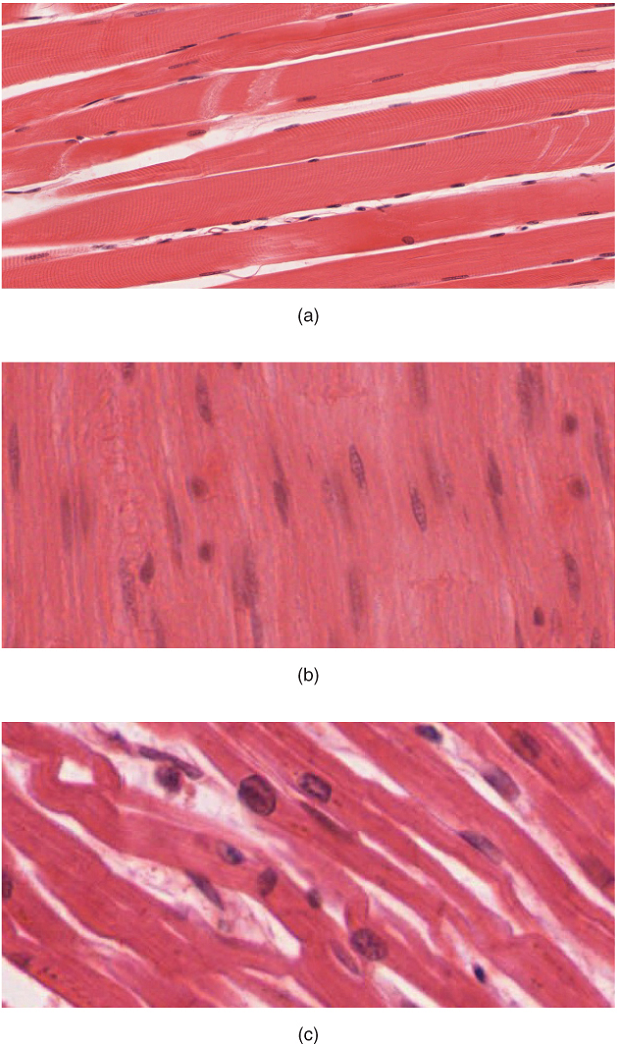
Differences among the three muscle types include the microscopic organization of their contractile proteins—actin and myosin. The actin and myosin proteins are arranged very regularly in the cytoplasm of individual muscle cells (referred to as fibers) in both skeletal muscle and cardiac muscle, which creates a pattern of stripes, called striations. The striations are visible with a light microscope under high magnification (Figure 13.1). Skeletal muscle fibers are multinucleated structures that compose the skeletal muscle. Cardiac muscle fibers each have one to two nuclei and are physically and electrically connected to each other so that the entire heart contracts as one unit (called a syncytium).
Because the actin and myosin are not arranged in such regular fashion in smooth muscle, the cytoplasm of a smooth muscle fiber (which has only a single nucleus) has a uniform, non-striated appearance (resulting in the name smooth muscle). However, the less organized appearance of smooth muscle should not be interpreted as less efficient.
Smooth muscle in the walls of arteries is a critical component that regulates blood pressure necessary to push blood through the circulatory system; and smooth muscle in the skin, visceral organs, and internal passageways is essential for moving all materials through the body.
Skeletal Muscle
The best-known feature of skeletal muscle is its ability to contract and cause movement. Skeletal muscles act not only to produce movement but also to stop movement, such as resisting gravity to maintain posture. Small, constant adjustments of the skeletal muscles are needed to hold a body upright or balanced in any position. Muscles also prevent excess movement of the bones and joints, maintaining skeletal stability and preventing skeletal structure damage or deformation. Joints can become misaligned or dislocated entirely by pulling on the associated bones; muscles work to keep joints stable. Skeletal muscles are located throughout the body at the openings of internal tracts to control the movement of various substances. These muscles allow functions, such as swallowing, urination, and defecation, to be under voluntary control. Skeletal muscles also protect internal organs (particularly abdominal and pelvic organs) by acting as a barrier or shield to external trauma and by supporting the weight of the organs.
Skeletal muscles contribute to the maintenance of homeostasis in the body by generating heat. Muscle contraction requires energy, and when ATP is broken down, heat is produced. This heat is very noticeable during exercise, when sustained muscle movement causes body temperature to rise, and in cases of extreme cold, when shivering produces random skeletal muscle contractions to generate heat.
Each skeletal muscle is an organ that consists of various integrated tissues. These tissues include the skeletal muscle fibers, blood vessels, nerve fibers, and connective tissue. Each skeletal muscle has three layers of connective tissue (called “mysia”) that enclose it and provide structure to the muscle as a whole, and also compartmentalize the muscle fibers within the muscle (Figure 13.2). Each muscle is wrapped in a sheath of dense, irregular connective tissue called the epimysium, which allows a muscle to contract and move powerfully while maintaining its structural integrity. The epimysium also separates muscle from other tissues and organs in the area, allowing the muscle to move independently.
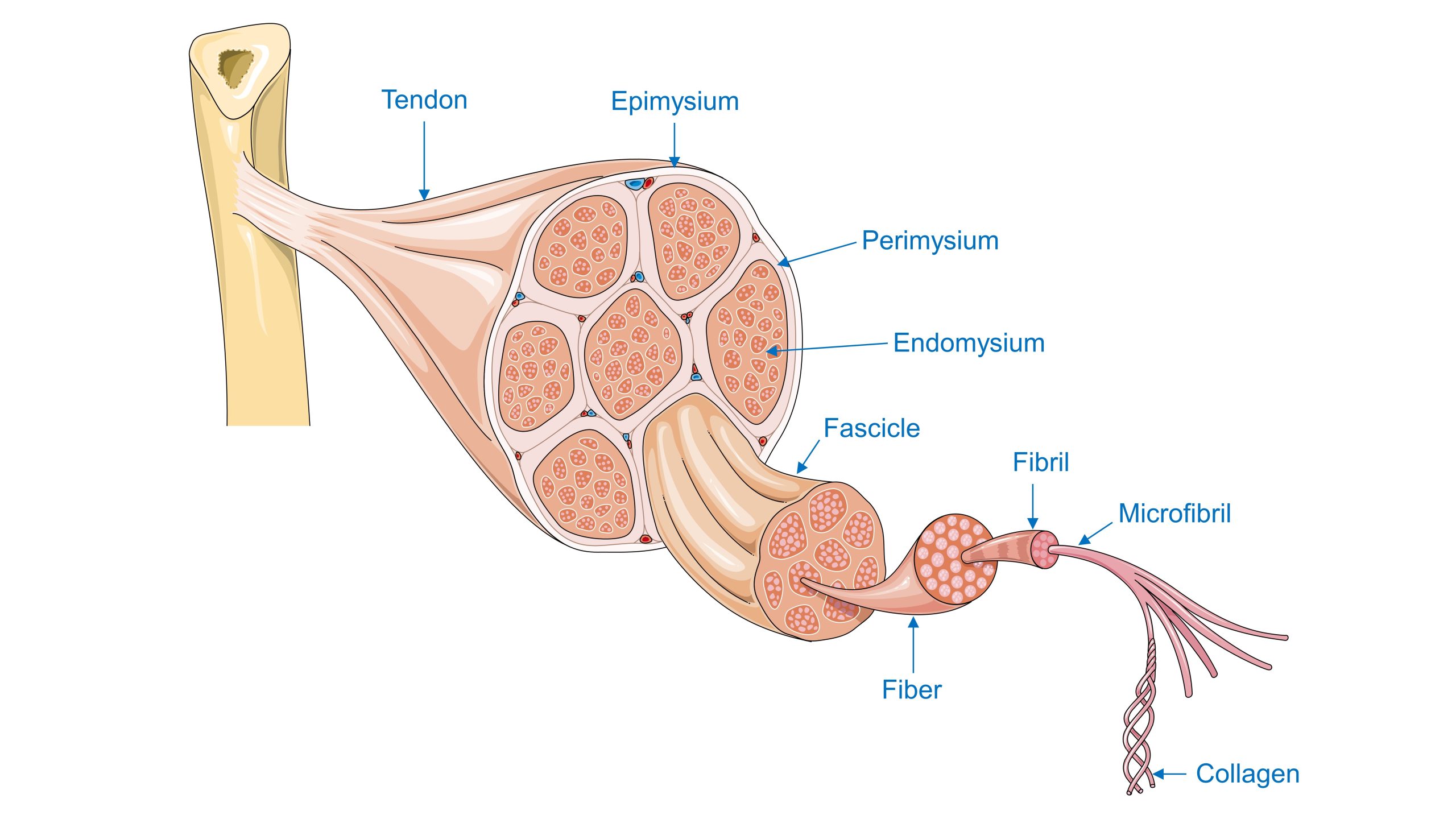
Inside each skeletal muscle, muscle fibers are organized into individual bundles, each called a fascicle, by a middle layer of connective tissue called the perimysium. This fascicular organization is common in muscles of the limbs; it allows the nervous system to trigger a specific movement of a muscle by activating a subset of muscle fibers within a bundle, or fascicle of the muscle. Inside each fascicle, each muscle fiber is encased in a thin connective tissue layer of collagen and reticular fibers called the endomysium. The endomysium contains the extracellular fluid and nutrients to support the muscle fiber. These nutrients are supplied via blood to the muscle tissue.
In skeletal muscles that work with tendons to pull on bones, the collagen in the three tissue layers (the mysia) intertwines with the collagen of a tendon. At the other end of the tendon, it fuses with the periosteum coating the bone. The tension created by the contraction of the muscle fibers is then transferred through the mysia, to the tendon, and then to the periosteum to pull on the bone for movement of the skeleton.
Every skeletal muscle is also richly supplied by blood vessels for nourishment, oxygen delivery, and waste removal. In addition, every muscle fiber in a skeletal muscle is innervated by the axon branch of a somatic motor neuron, which signals the fiber to contract. Unlike cardiac and smooth muscle, the only way to functionally contract a skeletal muscle is through signaling from the nervous system.
Skeletal Muscle Fibers
Because skeletal muscle cells are long and cylindrical, they are commonly referred to as muscle fibers. Skeletal muscle fibers can be quite large for human cells, with diameters up to 100 μm and lengths up to 30 cm (11.8 in) in the sartorius of the upper leg. During early development, embryonic myoblasts, each with its own nucleus, fuse with up to hundreds of other myoblasts to form the multinucleated skeletal muscle fibers. Multiple nuclei mean multiple copies of genes, permitting the production of the large amounts of proteins and enzymes needed for muscle contraction.
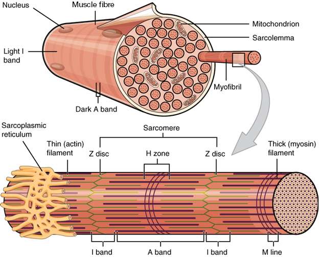
Some other terminology associated with muscle fibers is rooted in the Greek sarco, which means “flesh.” The plasma membrane of muscle fibers is called the sarcolemma, the cytoplasm is referred to as sarcoplasm, and the specialized smooth endoplasmic reticulum, which stores, releases, and retrieves calcium ions (Ca++) is called the sarcoplasmic reticulum (SR) (Figure 13.3). As will soon be described, the functional unit of a skeletal muscle fiber is the sarcomere, a highly organized arrangement of the contractile myofilaments actin (thin filament) and myosin (thick filament), along with other support proteins.
The Sarcomere
The Sarcomere: The striated appearance of skeletal muscle fibers is due to the arrangement of the myofilaments of actin and myosin in sequential order from one end of the muscle fiber to the other. Each packet of these microfilaments and their regulatory proteins, troponin and tropomyosin (along with other proteins) is called a sarcomere.
You can also watch the above video to learn more about macro- and microstructures of skeletal muscles by clicking the link or scanning the QR code below.

You can also watch the above Crash Course video to find out more about skeletal muscle structure by clicking the link or scanning the QR code below!

The sarcomere is the functional unit of the muscle fiber. The sarcomere itself is bundled within the myofibril that runs the entire length of the muscle fiber and attaches to the sarcolemma at its end. As myofibrils contract, the entire muscle cell contracts. Because myofibrils are only approximately 1.2 μm in diameter, hundreds to thousands (each with thousands of sarcomeres) can be found inside one muscle fiber.
Each sarcomere is around 2 μm in length with a cylinder-like arrangement and is bordered by structures called Z-discs (also called Z lines, because pictures are two-dimensional), to which the actin myofilaments are anchored (Figure 13.4). Because the actin and its troponin-tropomyosin complex (projecting from the Z-discs toward the center of the sarcomere) form strands that are thinner than the myosin, it is called the thin filament of the sarcomere. Likewise, because the myosin strands and their multiple heads (projecting from the center of the sarcomere, toward but not all the way to, the Z-discs) have more mass and are thicker, they are called the thick filament of the sarcomere.

Test Your Knowledge
- Describe the levels of muscle organization: fascia, fascicles, muscle fibers.
- Describe the following structures of a muscle cell: sarcolemma, sarcoplasm, nuclei, mitochondria, sarcoplasmic reticulum, transverse tubules, myofibrils, myofilaments, sarcomere.
- Define the following terms:
- Fascia
- Fascicle
- Epimysium
- Perimysium
- Endomysium
- Muscle fiber
- Muscle cell
- Tendon
- Explain why skeletal muscle fibers appear to have striations.
- Describe the location and general structure of each of the following:
- Sarcolemma
- Sarcoplasm
- Sarcoplasmic reticulum
- Sarcomere
- Myofilaments
- Myofibrils
- Transverse tubules
- Describe the following structures of a sarcomere: Z line, I band, A band, H zone, M line.
- Draw and fully label a diagram showing two adjacent, relaxed sarcomeres. Your diagram must include (labeled!):
- Z line
- I band
- A band
- H zone
- M line
- Sarcomere width
- Draw and fully label a diagram showing one fully contracted sarcomere. Your diagram must include labels:
- Z line
- I band
- A band
- H zone
- M line
- Sarcomere width
- Describe the basic structure of the thick and thin filaments and their primary protein components.
- Draw a diagram showing and identifying the major structural components of:
- A single myosin molecule
- A single thick filament
- A single thin filament
Region of the sacrum, bone forming the back part of the pelvic cavity.
Central nervous system fibers carrying sensory information from the spinal cord or periphery to the brain.
Central nervous system fibers carrying sensory information from the spinal cord or periphery to the brain.
Protein that makes up most of the thin myofilaments in a sarcomere muscle fibre.
Protein that makes up most of the thick cylindrical myofilament within a sarcomere muscle fiber.
Usually attached to bone, under voluntary control, each cell is a fiber that is multinucleated and striated.
Heart muscle, under involuntary control, composed of striated cells that attach to form fibres, each cell contains a single nucleus, contracts autonomously.
A multinucleate cell formed by the fusion of multiple cells or the division of nuclei.
Under involuntary control, moves internal organs, cells contain a single nucleus, are spindle-shaped, and do not appear striated; each cell is a fiber.
Steady state of body systems that living organisms maintain.
Nucleotide containing ribose and an adenine base that is essential in energy transfer.
Type of tissue that serves to hold in place, connect, and integrate the body’s organs and systems.
Central nervous system fibers carrying motor commands from the brain to the spinal cord or periphery.
Filaments between the arachnoid and pia mater within the subarachnoid space.
Filaments between the arachnoid and pia mater within the subarachnoid space.
The most abundant of three protein fibres found in the extracellular matrix of connective tissues.
Fine fibrous protein, made of collagen subunits, which cross-link to form supporting “nets” within connective tissue.
Thin, innermost membrane of the meninges that directly covers the surface of the CNS.
Fluid outside cells (plasma or interstitial fluid).
Mouth
Beginning of the large intestine, forming a small pouch.
Single process of the neuron that carries an electrical signal (action potential) away from the cell body toward a target cell.
Functional division of the nervous system that is concerned with conscious perception, voluntary movement, and skeletal muscle reflexes.
Four muscles that extend and stabilize the knee.
Thin, innermost membrane of the meninges that directly covers the surface of the CNS.
Remnants of the hollow center of the neural tube that are spaces for cerebrospinal fluid to circulate through the brain.
Four muscles that extend and stabilize the knee.
Remnants of the hollow center of the neural tube that are spaces for cerebrospinal fluid to circulate through the brain.
Specialized structures containing ependymal cells lining blood capillaries that filter blood to produce CSF in the four ventricles of the brain.
Specialized structures containing ependymal cells lining blood capillaries that filter blood to produce CSF in the four ventricles of the brain.
Portions of the ventricular system that are in the region of the cerebrum.
Portions of the ventricular system that are in the region of the cerebrum.
