29 Muscular System Organization
Unit Outline
The Muscular System
Think about the things that you do each day—talking, walking, sitting, standing, and running. All of these activities require the movement of particular skeletal muscles. Skeletal muscles are even used during sleep. The diaphragm is a sheet of skeletal muscle that has to contract and relax for you to breathe day and night. If you recall from your study of the skeletal system and joints, body movement occurs around the joints in the body. The focus of this chapter is on skeletal muscle organization. The system to name skeletal muscles will be explained; in some cases, the muscle is named by its shape, and in other cases, it is named by its location or attachments to the skeleton. If you understand the meaning of the name of the muscle, often it will help you remember its location and/or function. This chapter also will describe how skeletal muscles are arranged to accomplish movement and how other muscles may assist or be arranged on the skeleton to resist or carry out the opposite movement. The actions of the skeletal muscles will be covered in a regional manner, working from the head down to the toes.
Naming Skeletal Muscles
The Greeks and Romans conducted the first studies done on the human body in Western culture. The educated class of subsequent societies studied Latin and Greek, and therefore, the early pioneers of anatomy applied Latin and Greek terminology or roots when they named the skeletal muscles. The large number of muscles in the body and unfamiliar words can make learning the names of the muscles in the body seem daunting, but understanding the etymology can help. Etymology is the study of how the root of a particular word entered a language and how the use of the word evolved over time. Taking the time to learn the root of the words is crucial to understanding the vocabulary of anatomy and physiology (see Appendix II). When you understand the names of muscles, it will help you remember where the muscles are located and what they do (Figure 13.7, Table 13.2, Table 13.3, Table 13.4). Pronunciation of words and terms will take a bit of time to master, but after you have some basic information, the correct names and pronunciations will become easier.
| Example | Word | Latin Root 1 | Latin Root 2 | Meaning | Translation |
|---|---|---|---|---|---|
| abductor digiti minimi | abductor | ab = away from | duct = to move | moves away from | A muscle that moves the little finger/toe away |
| digiti | digitus = digit | refers to a finger or toe | |||
| minimi | minimus = minimal, tiny | little | |||
| adductor digiti minimi | adductor | ad = toward | duct = to move | moves toward | A muscle that moves the little finger/toe toward |
| digiti | digitus = digit | refers to a finger or toe | |||
| minimi | minimus = minimal, tiny | little |
Anatomists name the skeletal muscles according to a number of criteria, each of which describes the muscle in some way. These include naming the muscle after its shape, the direction of its muscle fibers, its size compared to other muscles in the area, its location in the body or the location of its attachments to the skeleton, how many origins it has, or its action. Often, a muscle’s name will refer to several of these characteristics (Table 13.3). You should be able to list the criteria and provide an example of each in the name of a muscle.
The shapes of some muscles are very distinctive, and the names, deltoid for the Greek letter delta (which looks like a triangle), reflect their shape. The direction of the muscle fibers and fascicles of a muscle can be used to name muscles by describing their orientation relative to the longitudinal axis of the body or of a limb, such as the rectus (straight) abdominis, or the oblique (at an angle) muscles of the abdomen.
For the buttocks, the size of the muscles influences the names: gluteus maximus (largest), gluteus medius (medium), and the gluteus minimus (smallest). Names are also given to muscles that indicate length—brevis (short) or longus (long). Some muscle names are used to indicate the number of muscles in a group. One example of this is the quadriceps, a group of four muscles located on the anterior (front) thigh.
The skeletal muscle’s anatomical location or its relationship to a particular bone often determines its name. Some muscles are named after their relative anatomical position: lateralis, medialis, dorsi (“dorsal”), anterior, and posterior.
The location of a muscle’s attachment can also appear in its name. When the name of a muscle is based on the attachments, the origin is always named first. For instance, the sternocleidomastoid muscle of the neck has a dual origin on the sternum (“sterno”) and clavicle (“cleido”) and inserts on the mastoid process of the temporal bone. Other muscle names can provide information as to how many origins a particular muscle has, such as the biceps brachii. The prefix bi- indicates that the muscle has two origins, and tri indicates three origins.
The last feature by which to name a muscle is its action. When muscles are named for the movement they produce, one can find action words in their name.
|
Criteria |
Descriptions |
Meaning |
Example |
|---|---|---|---|
|
Shape |
Orbicularis |
Orbit (ring) |
Orbicularis oculi |
|
Deltoid |
Triangle |
Deltoid |
|
|
Orientation |
Rectus |
Straight |
Rectus femoris |
|
Oblique |
At an angle |
Abdominis external oblique |
|
|
Size |
Brevis |
Short |
Adductor brevis |
|
Longissimus/longus |
Long |
Adductor longus |
|
|
Maximus |
Largest |
Gluteus maximus |
|
|
Medius |
Medium |
Gluteus medius |
|
|
Minimus |
Smallest |
Gluteus minimus |
|
|
Anatomical position |
Medialis |
Medial (along the midline) |
Vastus medialis |
|
Lateralis |
Lateral (away from midline) |
Vastus lateralis |
|
|
Dorsi |
Dorsal (back) |
Latissimus dorsi |
|
|
Anterior |
Forward |
Tibialis anterior |
|
|
Posterior |
Rear |
Tibialis posterior |
|
|
Abdominis |
abdomen |
Abdominis external oblique |
|
|
Bone name |
various |
Rectus femoris (along femur) Frontalis (on top of frontal bone) |
|
|
Number of origins |
Bi-, tri- |
|
Biceps brachii |
|
Origin/Insertion location on skeleton |
Names of bones or parts of bones |
various |
Sternocleidomastoid (origins: sternum, clavicle; insertion: mastoid process) |
|
Actions |
Muscle actions |
|
Adductor longus |
| Example | Latin or Greek Translation | Mnemonic Device |
|---|---|---|
| ad- | to; toward | ADvance toward your goal |
| ab- | away from | Aliens ABduct you away from home |
| sub- | under | SUBmarines move underwater |
| -ductor | something that moves | A conDUCTOR makes a train move |
| anti- | against | If you are ANTIsocial, you are against engaging in social activities |
| epi- | on top of | She is the EPItome of goodness |
| apo- | to the side of | An APOstrophe separates parts of a contraction from each other |
| longissimus | longest | “Longissimus” is longer than the word “long” |
| longus | long | LONGus |
| brevis | short | BRief |
| maximus | large | MAXIMUm size |
| minimus | tiny; little | MINIMUm size |
| medius | between large and tiny | Of MEDIUm size |
| rectus | straight | A situation is considered RECTified when it is straightened out |
| multi | many | A rainbow is MULTIcoloured |
| uni- | one | A UNIcorn has one horn |
| bi- (Latin root) or di- (Greek root) | two | You can DIvide something into two pieces; BIcycles have two wheels |
| tri- | three | To TRIple your money, you multiply it by three |
| quad- | four | QUADruplets are four children born at one birth |
| externus | outside | EXTERNal |
| internus | inside | INTERNal |
Axial Muscles of the Head, Neck, and Back
The skeletal muscles are divided into two categories: axial (muscles of the trunk and head) and appendicular (muscles of the arms and legs). This system reflects the bones of the skeleton system, which are also arranged in this manner. The axial muscles are grouped based on location, function, or both. Some of the axial muscles may seem to blur the boundaries because they cross over to the appendicular skeleton. The first grouping of the axial muscles you will review includes the muscles of the head and neck, then you will review the muscles of the vertebral column, and finally you will review the oblique and rectus muscles.
Muscles That Move the Head: The head, attached to the top of the vertebral column, is balanced, moved, and rotated by the neck muscles. When these muscles act unilaterally, the head rotates. When they contract bilaterally, the head flexes or extends. The major muscle that laterally flexes and rotates the head is the sternocleidomastoid. In addition, both muscles working together are the flexors of the head. Place your fingers on both sides of the neck and turn your head to the left and to the right. You will feel the movement originate there. This muscle divides the neck into anterior and posterior triangles when viewed from the side (Figure 13.8).
Muscles of the Posterior Neck and the Back: The posterior muscles of the neck are primarily concerned with head movements, like extension. The back muscles stabilize and move the vertebral column and are grouped according to the lengths and direction of the fascicles.
The erector spinae group forms the majority of the muscle mass of the back, and it is the primary extensor of the vertebral column. It controls flexion, lateral flexion, and rotation of the vertebral column and maintains the lumbar curve. The erector spinae comprises the iliocostalis (laterally placed) group, the longissimus (intermediately placed) group, and the spinalis (medially placed) group.
Muscles of the Abdominal Wall and Thorax
It is a complex job to balance the body on two feet and walk upright. The muscles of the vertebral column, thorax, and abdominal wall extend, flex, and stabilize different parts of the body’s trunk. The deep muscles of the core of the body help maintain posture as well as carry out other functions. The brain sends out electrical impulses to these various muscle groups to control posture by alternate contraction and relaxation. This is necessary so that no single muscle group becomes fatigued too quickly. If any one group fails to function, body posture will be compromised.
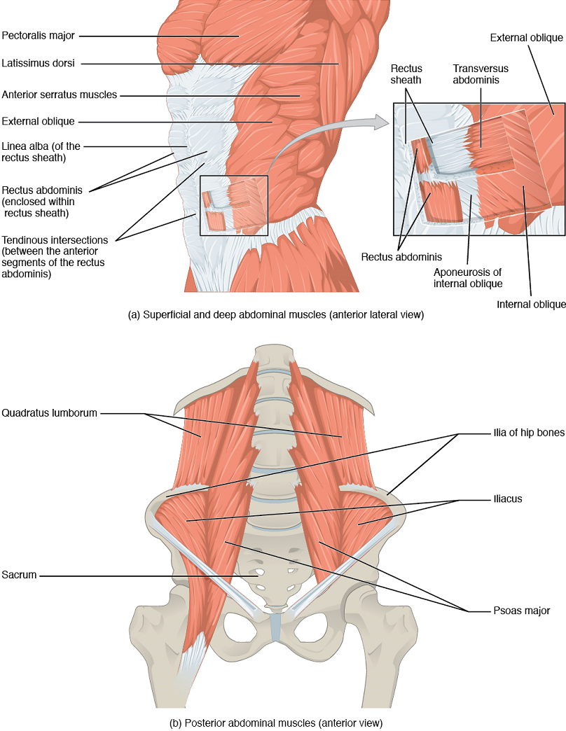
Muscles of the Abdomen: There are four pairs of abdominal muscles that cover the anterior and lateral abdominal region and meet at the anterior midline. These muscles of the anterolateral abdominal wall can be divided into four groups: the external obliques, the internal obliques, the transversus abdominis, and the rectus abdominis (Figure 13.10).
The external obliques, internal obliques, and transversus abdominis are three flat skeletal muscles in the antero-lateral wall of the abdomen. This arrangement of three bands of muscles in different orientations allows various movements and rotations of the trunk. The three layers of muscle also help to protect the internal abdominal organs in an area where there is no bone.
The linea alba is a white fibrous band that is made of the bilateral rectus sheaths that join at the anterior midline of the body. These enclose the rectus abdominis muscles (a pair of long, linear muscles, commonly called the “sit-up” muscles) that originate at the pubic crest and symphysis and extend the length of the body’s trunk to insert on the sternum and ribs five to seven. These muscles flex the abdomen, as in the motion or bending forward or doing a sit-up exercise. Each muscle is segmented by three transverse bands of collagen fibers called the tendinous intersections. This results in the look of “six-pack abs,” as each segment hypertrophies on individuals at the gym who do many sit-ups.
The posterior abdominal wall is formed by the lumbar vertebrae, parts of the ilia of the hip bones, psoas major and iliacus muscles, and quadratus lumborum muscle. This part of the core plays a key role in stabilizing the rest of the body and maintaining posture.
Muscles of the Thorax: The muscles of the chest facilitate breathing by changing the size of the thoracic cavity. When you inhale, your chest rises because the cavity expands. Alternately, when you exhale, your chest falls because the thoracic cavity decreases in size.
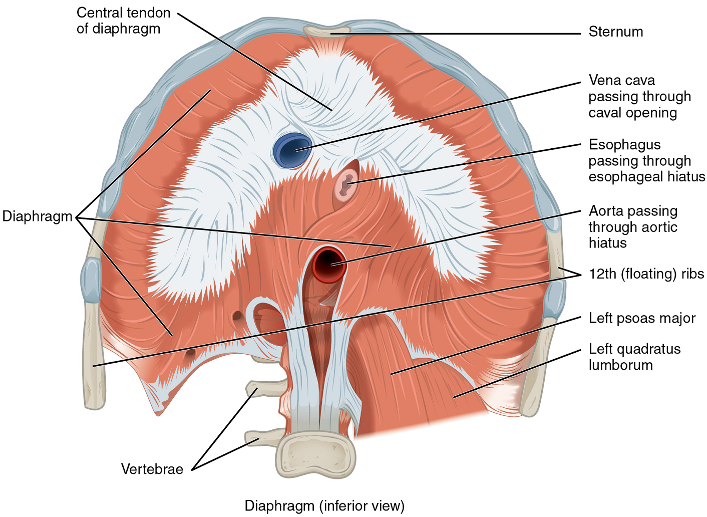
The Diaphragm: The change in volume of the thoracic cavity during breathing is due to the alternate contraction and relaxation of the diaphragm (Figure 13.11). It separates the thoracic and abdominal cavities, and is dome-shaped at rest. The superior surface of the diaphragm is convex, creating the elevated floor of the thoracic cavity. The inferior surface is concave, creating the curved roof of the abdominal cavity.
Defecating, urination, and even childbirth involve cooperation between the diaphragm and abdominal muscles (this cooperation is referred to as the “Valsalva maneuver”). You hold your breath by a steady contraction of the diaphragm; this stabilizes the volume and pressure of the peritoneal cavity. When the abdominal muscles contract, the pressure cannot push the diaphragm up, so it increases pressure on the intestinal tract (defecation), urinary tract (urination), or reproductive tract (childbirth).
The inferior surface of the pericardial sac and the inferior surfaces of the pleural membranes (parietal pleura) fuse onto the central tendon of the diaphragm. To the sides of the tendon are the skeletal muscle portions of the diaphragm, which insert into the tendon while having a number of origins including the xiphoid process of the sternum anteriorly, the inferior six ribs and their cartilages laterally, and the lumbar vertebrae and 12th ribs posteriorly.
The diaphragm also includes three openings for the passage of structures between the thorax and the abdomen. The inferior vena cava passes through the caval opening, and the esophagus and attached nerves pass through the esophageal hiatus. The aorta, thoracic duct, and azygous vein pass through the aortic hiatus of the posterior diaphragm.
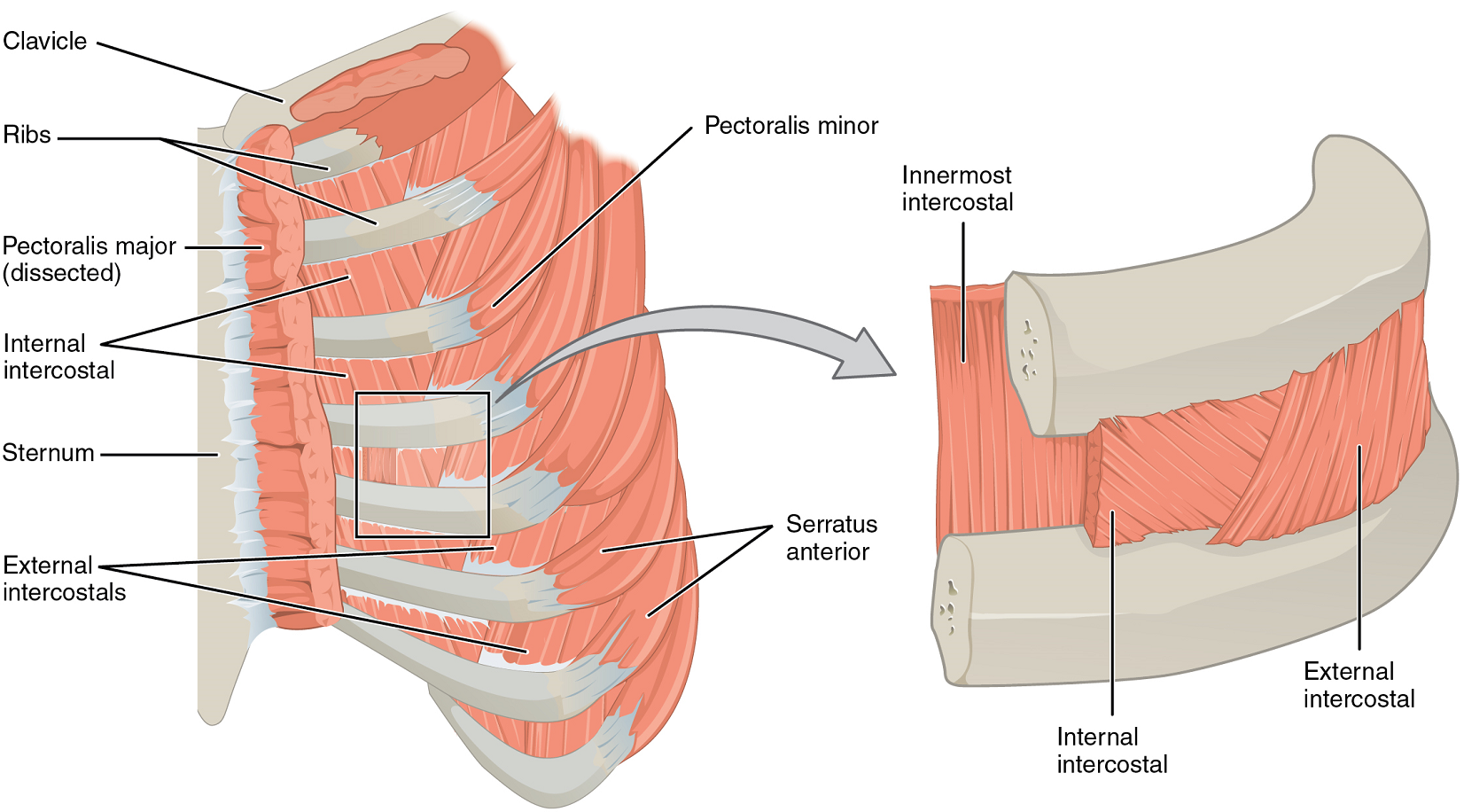
The Intercostal Muscles: There are three sets of muscles, called intercostal muscles, which span each of the intercostal spaces. The principal role of the intercostal muscles is to assist in breathing by changing the dimensions of the rib cage (Figure 13.12).
The 11 pairs of superficial external intercostal muscles aid in inspiration of air during breathing because when they contract, they raise the rib cage, which expands it. The 11 pairs of internal intercostal muscles, just under the externals, are used for expiration because they draw the ribs together to constrict the rib cage. The innermost intercostal muscles are the deepest, and they act as synergists for the action of the internal intercostals.
Muscles of the Pectoral Girdle and Upper Limbs
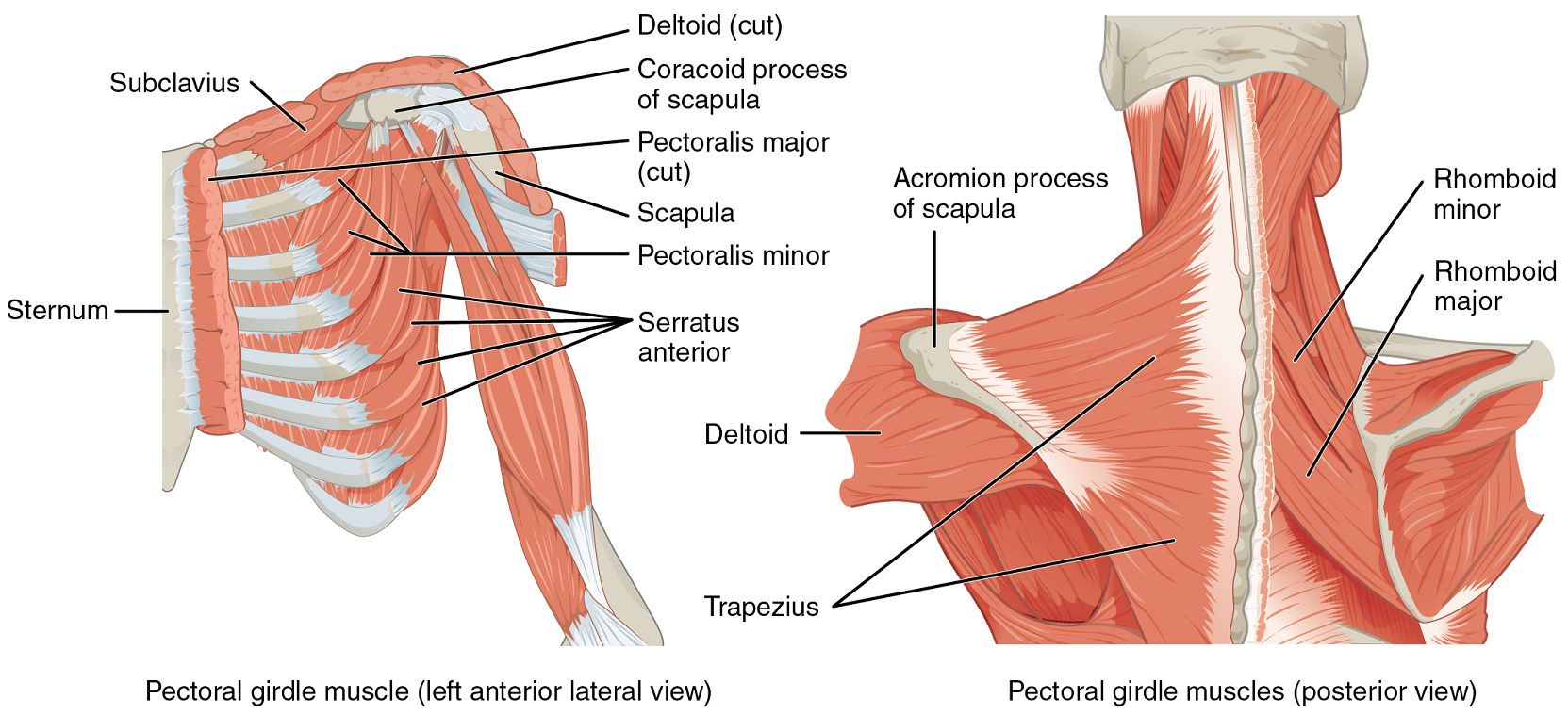
Muscles of the shoulder and upper limb can be divided into four groups: muscles that stabilize and position the pectoral girdle, muscles that move the entire arm, muscles that move the forearm, and muscles that move the wrists, hands, and fingers. The pectoral girdle, or shoulder girdle, consists of the lateral ends of the clavicle and scapula, along with the proximal end of the humerus, and the muscles covering these three bones to stabilize the shoulder joint. The girdle creates a base from which the head of the humerus, in its ball-and-socket joint with the glenoid fossa of the scapula, can move the arm in multiple directions.
Muscles that position the pectoral girdle are located either on the anterior thorax or on the posterior thorax (Figure 13.13). Among the most important of these is the trapezius, located in the posterior thorax that originate on the skull and upper vertebral column and insert on the clavicle and scapula. The trapezius is capable of diverse movements such as elevation and depression of the scapula (shrugging shoulders), moving the scapula together, and tilting the head backward.
Muscles that move the humerus: Similar to the muscles that position the pectoral girdle, muscles that cross the shoulder joint and move the humerus bone of the arm include both axial and scapular muscles (Figure 13.14 and Table 13.5). The two axial muscles are the pectoralis major and the latissimus dorsi. The pectoralis major is thick and fan-shaped, covering much of the superior portion of the anterior thorax. The broad, triangular latissimus dorsi is located on the inferior part of the back, where it inserts into a thick connective tissue sheath called an aponeurosis.
The rest of the shoulder muscles originate on the scapula. The anatomical and ligamental structure of the shoulder joint and the arrangements of the muscles covering it allow the arm to carry out different types of movements. The deltoid, the thick muscle that creates the rounded lines of the shoulder, is the major abductor of the arm, but it also facilitates flexing and medial rotation, as well as extension and lateral rotation. Named for its location, the supraspinatus (superior to the spine of the scapula) abducts the arm. The thick and flat teres major extends the arm and assists in adduction and medial rotation of it. The long teres minor laterally rotates and extends the arm. Finally, the coracobrachialis flexes and adducts the arm (Table 13.5)
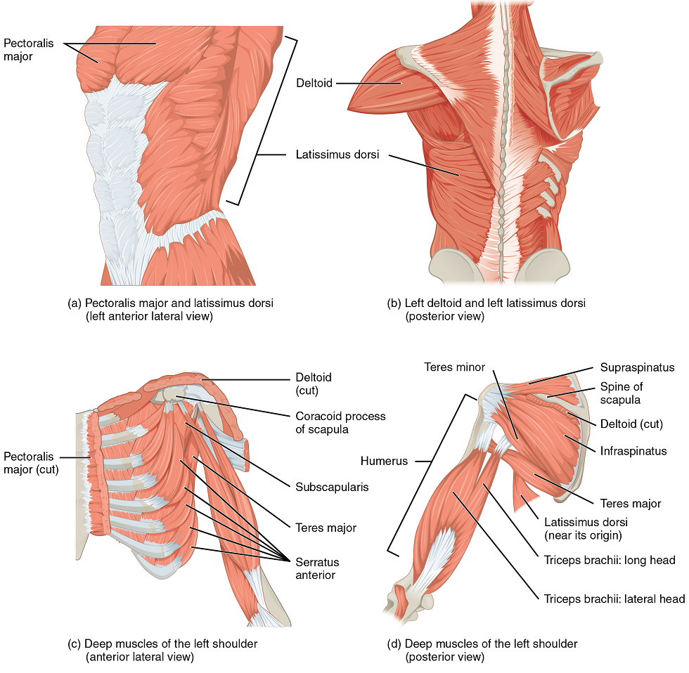
The tendons of the deep subscapularis, supraspinatus, infraspinatus, and teres minor connect the scapula to the humerus, forming the rotator cuff (musculotendinous cuff), the circle of tendons around the shoulder joint. When baseball pitchers undergo shoulder surgery, it is usually on the rotator cuff, which becomes pinched and inflamed and may tear away from the bone due to the repetitive motion of bringing the arm overhead to throw a pitch.
| Movement | Target | Target motion direction | Prime mover | Origin | Insertion |
|---|---|---|---|---|---|
| Axial muscles | |||||
|
Brings elbows together; moves elbow up (as during an uppercut punch) |
Humerus |
Flexion; adduction; medial rotation |
Pectoralis major |
Clavicle, sternum, cartilage of certain ribs (1–6 or 1–7); aponeurosis of external oblique muscle |
Greater tubercle of humerus |
|
Moves elbow back (as in elbowing someone behind you); spreads elbows apart |
Humerus, scapula |
Humerus:extension, adduction and medial rotation;
Scapula: depression |
Latissimus dorsi | Thoracic vertebrae (T7–T12); Lumbar vertebrae; lower ribs (9–12); iliac crest | Intertubercular sulcus of humerus |
| Scapular muscles | |||||
| Lifts arms at shoulder | Humerus | Abduction; flexion; extension; medial and lateral rotation | Deltoid | Trapezius; Clavicle; Acromion spine of scapula | Deltoid tuberosity of humerus |
| Rotates elbow outwards, as during a tennis swing | Humerus | Abduction | Supraspinatus | Supraspinous fossa of scapula | Greater tubercle of humerus |
| Rotate elbow outward | Humerus | Extension; Adduction | Teres major | Posterior surface of scapula | Intertubercular sulcus of humerus |
| Moves elbow up and across body, as when putting hand on chest. | Humerus | Flexion; adduction | Coracobrachialis | Coracoid process of scapula | Medial surface of humerus shaft |
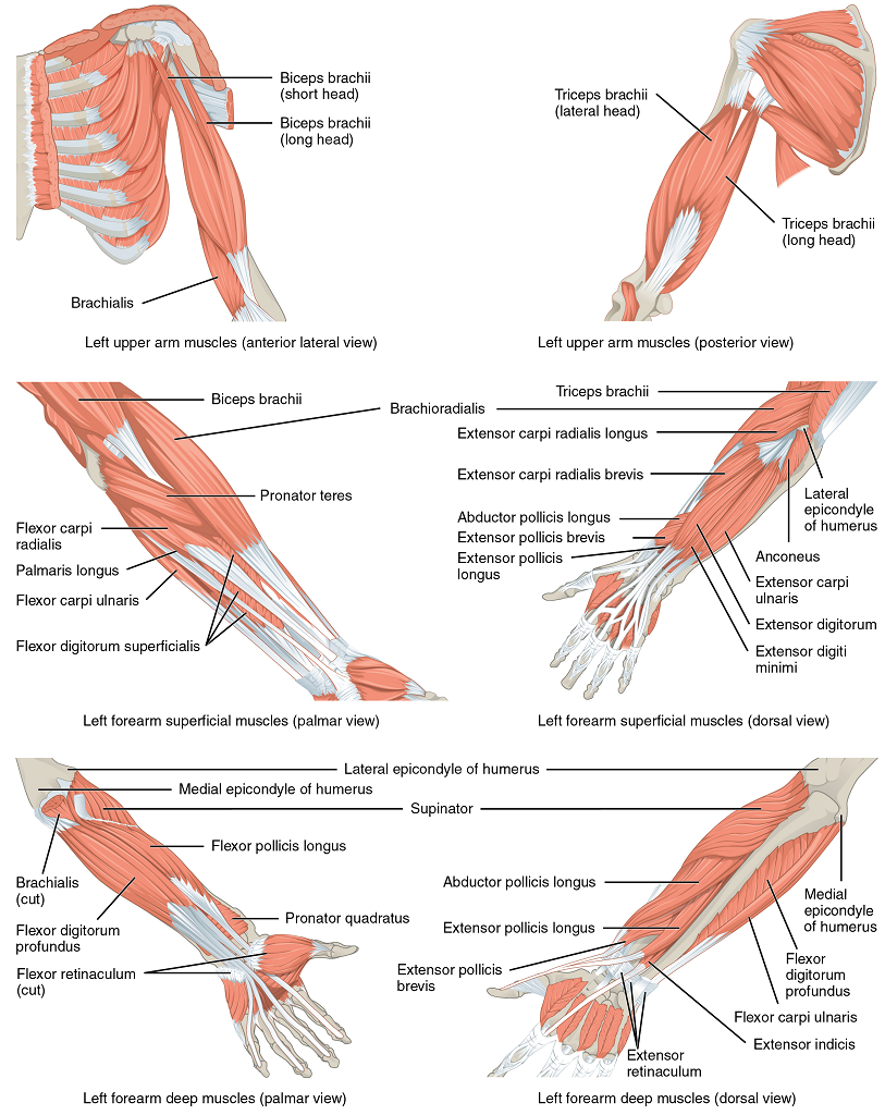
Muscles That Move the Forearm: The forearm, made of the radius and ulna bones, has four main types of action at the hinge of the elbow joint: flexion, extension, pronation, and supination. The forearm flexors include the biceps brachii and brachioradialis. The major extensor is the triceps brachii.
The biceps brachii and brachioradialis flex the forearm. The two-headed biceps brachii crosses the shoulder and elbow joints to flex the forearm, also taking part in supinating the forearm at the radioulnar joints and flexing the arm at the shoulder joint. The brachioradialis can flex the forearm quickly or help lift a load slowly. These muscles and their associated blood vessels and nerves form the anterior compartment of the arm (anterior flexor compartment of the arm; Figure 13.15 and Table 13.6).
| Movement | Target | Target motion direction | Prime mover | Origin | Insertion |
| Anterior muscles (flexion) | |||||
|
Performs a bicep curl; also allows palm of hand to point toward body while flexing |
Forearm |
Flexion; supination |
Biceps brachii |
Scapula: coracoid process and tubercle above glenoid cavity |
Radial tuberosity |
|
Assists and stabilizes elbow while performing a bicep curl |
Forearm |
Flexion | Brachioradialis | Lateral supracondylar ridge at distal end of humerus | Base of styloid process of radius |
| Posterior muscle (extension) | |||||
| Extends forearm, as during a punch | Forearm | Extension | Triceps brachii | Infraglenoid tubercle of scapula; posterior shaft of humerus; posterior humeral shaft distal to radial groove | Olecranon process of ulna |
Appendicular Muscles of the Pelvic Girdle and Lower Limbs
The appendicular muscles of the lower body position and stabilize the pelvic girdle, which serves as a foundation for the lower limbs. Comparatively, there is much more movement at the pectoral girdle than at the pelvic girdle. There is very little movement of the pelvic girdle because of its connection with the sacrum at the base of the axial skeleton. The pelvic girdle has less range of motion because its function is to stabilize and support the body.
Muscles of the Thigh: What would happen if the pelvic girdle, which attaches the lower limbs to the torso, were capable of the same range of motion as the pectoral girdle? For one thing, walking would expend more energy if the heads of the femurs were not secured in the acetabula of the pelvis. The body’s center of gravity is in the area of the pelvis. If the center of gravity were not to remain fixed, standing up would be difficult as well. Therefore, what the leg muscles lack in range of motion and versatility, they make up for in size and power, facilitating the body’s stabilization, posture, and movement.
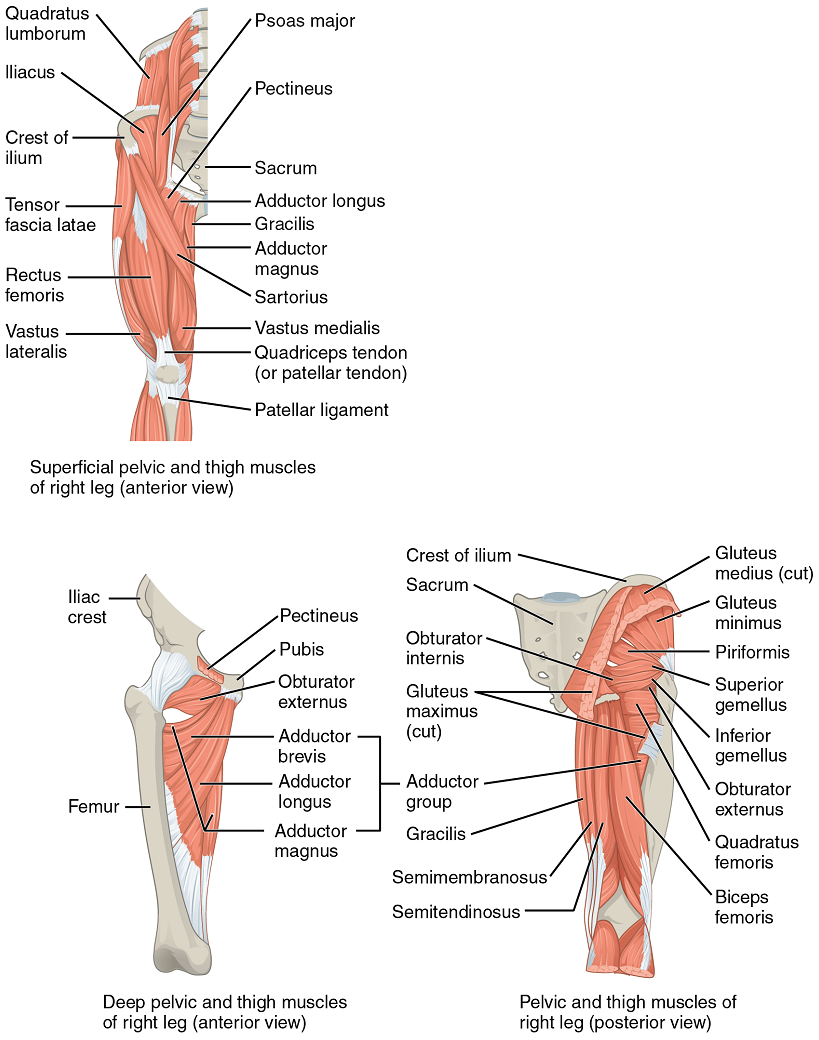
Gluteal Region Muscles That Move the Femur: Most muscles that insert on the femur (the thigh bone) and move it originate on the pelvic girdle. The psoas major and iliacus make up the iliopsoas group. Some of the largest and most powerful muscles in the body are the gluteal muscles or gluteal group. The gluteus maximus is the largest and deep to the gluteus maximus is the gluteus medius (Figure 13.16 and Table 13.7).
| Movement | Target | Target motion direction | Prime mover | Origin | Insertion |
| Iliopsoas group | |||||
|
Raises knee at hip, as if performing a knee attack; assists lateral rotators in twisting thigh (and lower leg) outward; assists with bending over, maintaining posture. |
Femur |
Thigh: flexion and lateral rotation; torso: flexion |
Psoas major Iliacus |
Psoas major: Lumbar vertebrae; and thoracic vertebra T12; Iliacus: iliac fossa, iliac crest and lateral sacrum |
Lesser trochanter of femur |
| Gluteal group | |||||
| Lowers knee and moves thigh back, as when getting ready to kick a ball | Femur | Extension | Gluteus maximus | Dorsal ilium; sacrum; coccyx | Gluteal tuberosity of femur; iliotibial tract |
| Opens thighs, as when doing a split | Femur | Abduction | Gluteus medius | Lateral surface of ilium | Greater trochanter of femur |
Thigh Muscles That Move the Femur, Tibia, and Fibula: Deep fascia in the thigh separates it into medial, anterior, and posterior compartments (Figure 13.16). The major muscle in the medial compartment of the thigh is the strap-like gracilis that adducts the thigh in addition to flexing the leg at the knee.
The muscles of the anterior compartment of the thigh flex the thigh and extend the leg. This compartment contains the quadriceps femoris group, which actually comprises four muscles that extend and stabilize the knee. The most important of these is the rectus femoris, located on the anterior aspect of the thigh. The tendon common to all four is the quadriceps tendon (patellar tendon), which inserts into the patella and continues below it as the patellar ligament. The patellar ligament attaches to the tibial tuberosity. In addition to the quadriceps femoris, the sartorius is a band-like muscle that extends from the anterior superior iliac spine to the medial side of the proximal tibia. This versatile muscle flexes the leg at the knee and flexes, abducts, and laterally rotates the leg at the hip. This muscle allows us to sit cross-legged.
The posterior compartment of the thigh includes muscles that flex the leg and extend the thigh. The three long muscles on the back of the knee are the hamstring group, which flexes the knee. These are the biceps femoris, semitendinosus, and semimembranosus. The tendons of these muscles form the popliteal fossa, the diamond-shaped space at the back of the knee.
Muscles That Move the Feet and Toes: Similar to the thigh muscles, the muscles of the leg are divided by deep fascia into compartments, although the leg has three: anterior, lateral, and posterior (Figure 13.17).
The muscles in the anterior compartment of the leg: the tibialis anterior, a long and thick muscle on the lateral surface of the tibia, the extensor hallucis longus, deep under it, and the extensor digitorum longus, lateral to it, all contribute to raising the front of the foot when they contract. The fibularis tertius, a small muscle that originates on the anterior surface of the fibula, is associated with the extensor digitorum longus and sometimes fused to it but is not present in all people. Thick bands of connective tissue called the superior extensor retinaculum (transverse ligament of the ankle) and the inferior extensor retinaculum hold the tendons of these muscles in place during dorsiflexion.
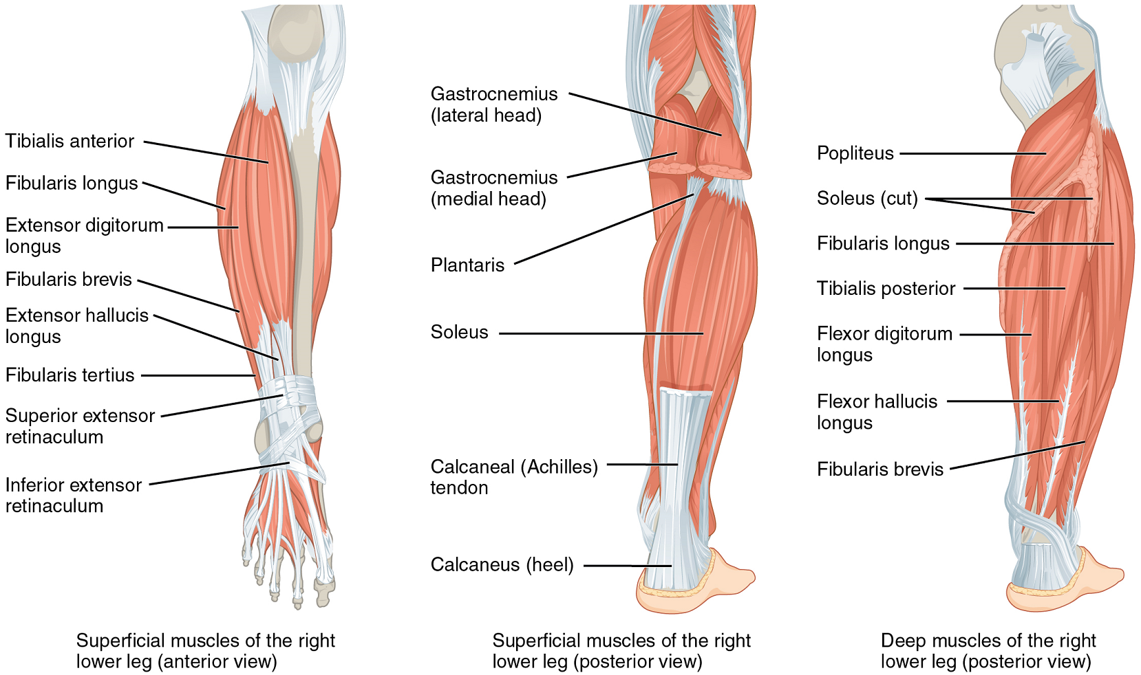
The lateral compartment of the leg includes two muscles: the fibularis longus (peroneus longus) and the fibularis brevis (peroneus brevis). The superficial muscles in the posterior compartment of the leg all insert onto the calcaneal tendon (Achilles tendon), a strong tendon that inserts into the calcaneal bone of the ankle. The muscles in this compartment are large and strong and keep humans upright. The most superficial and visible muscle of the calf is the gastrocnemius. Deep to the gastrocnemius is the wide, flat soleus. The plantaris runs obliquely between the two; some people may have two of these muscles, whereas no plantaris is observed in about seven percent of other cadaver dissections. The plantaris tendon is a desirable substitute for the fascia lata in hernia repair, tendon transplants, and repair of ligaments. There are four deep muscles in the posterior compartment of the leg as well: the popliteus, flexor digitorum longus, flexor hallucis longus, and tibialis posterior.
Practice
For the activity below, click the correct answer to each question.
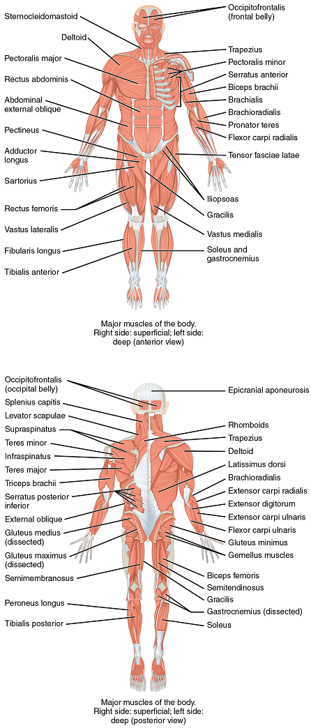
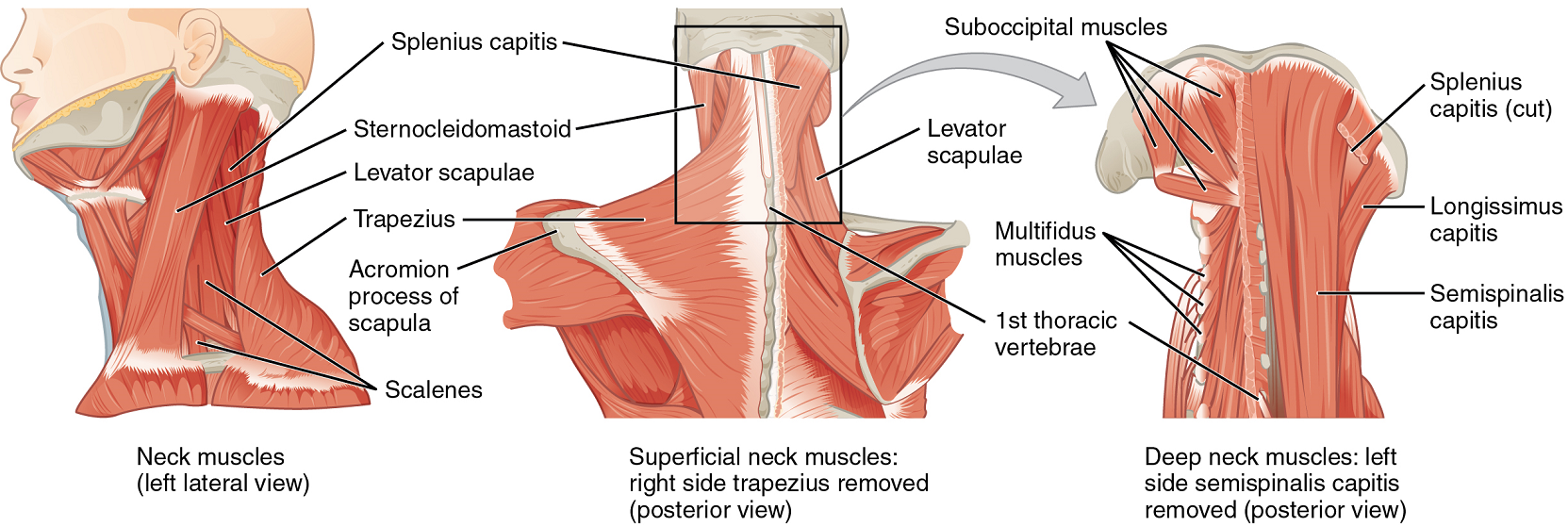
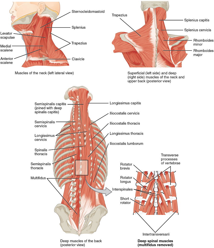
Image Descriptions

Figure 13.1 image description: A comparison of three micrographs of the primary muscle tissues types: skeletal, smooth, and cardiac. All tissues were examined using a light microscope (LM) at the same magnification: 1600X. Skeletal and cardiac muscle are long and have prominent striations that are lacking in smooth muscle. Skeletal muscle fibers are arranged in parallel to one another, while cardiac muscle fibers are branched and interwoven like a basket. Smooth muscle cells are the smallest and least distinct of the three and have a delicate spindle shape, meaning they have tapered ends on both sides. [Return to image.]

Figure 13.2 image description: An illustration of the nested layers of connective tissues that form a skeletal muscle. A large muscle resembling a gastrocnemius connects to a bone via a tendon that is an extension of the outermost covering called the epimysium. A cross section reveals that the muscle is composed of multiple bundles called fascicles that are collectively bound together by the epimysium. Each fascicle is in turn wrapped by a layer called the perimysium. Inside the fascicle are the individual skeletal muscle cells, called fibers. Each fiber is enclosed by a layer called endomysium. Finally, each fiber is composed of fibrils. Contractile elements of the muscle fibers are the actin and myosin but not labeled.. [Return to image.]

Figure 13.3 image description: A muscle fiber/cell is composed internally of many myofibrils and multiple nuclei externally. The entire muscle fiber is covered by a layer called the sarcolemma. Mitochondria are located amidst the microfibrils. The microfibrils are surrounded by a net-like sarcoplasmic reticulum and internally are composed of many thin (actin) and thick (myosin) filaments. These filaments are differentially arranged into specific regions of a contractile unit called a sarcomere. [Return to image.]

Figure 13.4 image description: A close-up representation of a sarcomere in muscle distinguishes the band/striations. The lighter/less-dense region, called the I Band, is formed exclusively by thin filaments (actin), while the darker/denser region called the A Band is filled with mostly thick filaments (myosin). A further close-up models myosin filaments as a thick rope sprouting multiple heads like golf clubs along its length. Each head is attached via a flexible neck and offers separate attachment regions for actin and myosin. On the other hand, the actin filament is modeled as a chain of bead-like elements containing a hollow binding site for the myosin head but surrounded by a thread-like molecule called tropomyosin. The tropomyosin appears to interfere with the access to the myosin binding location on the actin. Another component, called troponin, is associated with the tropomyosin but is not abundant. [Return to image.]

Figure 13.5 image description: This multi-part image shows different types of movements that are possible by different joints in the body. Labels read (from top, left): a and b angular movements: flexion and extension at the shoulders and knees; c) angular movements: flexion and extension of the neck (arrows pointing left and right to indicate movement). Labels (from bottom, left) read: d) angular movements: flexion and extension of the vertical column; e) angular movements abduction, adduction, and circumduction of the upper limb at the shoulder; f) rotation of the head, neck, and lower limb. [Return to image.]

Figure 13.6 image description: This multi-part image shows different types of movements that are possible by different joints in the body. Labels read (from top, left): g) pronation (P) and supination (S), h) dorsiflexion and plantar flexion, i) inversion and eversion. Labels read (from bottom, left): j) protraction and retraction, k) elevation and depression, and l) opposition. [Return to image.]

Figure 13.7 image description: This two-part diagram shows anterior (upper diagram) and posterior (lower diagram) views of the superficial muscle system.  In the anterior view, different muscles are identified on the left and right side of the body, although all muscles are found on each side. Labels on the right side of the body read (from top to bottom): sternocleidomastoid, deltoid, pectoralis major, rectus abdominis, abdominal external oblique, pectineus, adductor longus, sartorius, rectus femoris, vastus lateralis, fibularis longus, and tibialis anterior. Labels for left side of the body read (from top to bottom): occipitofrontalis (frontal belly), trapezius, pectoralis minor, serratus anterior, biceps brachii, brachialis, brachioradialis, pronator teres, flexor carpi radialis, tensor fasciae latae, iliopsoas, gracilis, vastus medialis, soleus, and and gastrocnemius. As with the anterior view of muscles, the posterior view has different muscles identified on the left and right sides. Labels on the left side of the body read (from top to bottom): occipitofrontalis (occipital belly), splenius capitis, levator scapulae, supraspinatus, teres minor, intraspinatus, teres major, triceps brachii, serratus posterior inferior, external oblique, gluteus medius (dissected), gluteus maximus (dissected) semimembranosus, peroneus longus, and tibialis posterior. Labels for right side of the body read (from top to bottom): epicranial aponeurosis, rhomboids, trapezius, deltoid, latissimus dorsi, brachioradialis, extensor carpi radialis, extensor digitorum, extensor carpi ulnaris, flexor carpi ulnaris, gluteus minimus, gemellus muscles, biceps femoris, semitendinosus, gracilis, gastrocnemius (dissected), and soleus. [Return to image.]

Figure 13.8 image description: This three-part diagram shows the superficial (left and middle diagram) and deep neck muscles (right diagram). In the leftmost diagram, the superficial neck muscles are labeled on an illustration of the left lateral view of a neck. Labels read (from top to bottom): splenius capitis, sternocleidomastoid, levator scapulae, trapezius, and scalenes. In the middle diagram, the superficial neck muscles are viewed additionally from posterior view but with the right trapezius muscle removed to reveal more of these muscles. Labels read (from top to bottom): splenius capitis, sternocleidomastoid, levator scapulae, acromion process of scapula, and 1st thoracic vertebrae. The rightmost diagram shows the deep neck muscles from the posterior view and the semispinalis capitis removed from the left side.  Labels read (from top to bottom): suboccipital muscles, splenius capitis (cut), longissimus capitis, semispinialis capitis, multifidus muscles, and 1st thoracic vertebrae. [Return to image.]

Figure 13.9 image description: This four-part diagram shows more neck muscles and back. The upper-left diagram shows the neck in the left lateral view. Labels read (from top to bottom): sternocleidomastoid, splenius, trapezius, levator scapulae, medial scalene, anterior scalene, clavicle. The upper-right diagram depicts the neck and upper back in posterior view; the trapezius muscle has been removed from the right side to show the deep neck muscles. Labels read (from top to bottom): splenius capitis, splenius cervicis, rhomboides minor, and rhomboides major. The bottom-left diagram shows the neck and back in posterior view. Deep muscle labels read (from top to bottom): semispinalis capitis (joined with deep spinalis capitis), longissimus capitis, semispinalis cervicis, iliocostalis cervicis, longissimus cervicis, illiocostalis thoracis, spinalis thoracis, longissimus thoracis, iliocostalis lumborum, and multifidus. The bottom-right diagram is an inset from the bottom-left diagram and shows the deepest spinal muscles with the multifidus removed. These muscle labels read (from top to bottom): rotator brevis, rotator longus, short rotator, interpspinales, and intertransversarii. [Return to image.]

Figure 13.10 image description: This two-part diagram shows anterior (upper diagram) and posterior (lower diagram) abdominal muscles. The anterior view labels read (from top to bottom): pectoralis major, latissimus dorsi, anterior serratus muscles, external oblique, linea alba (of the rectus sheath), rectus abdominis (enclosed within rectus sheath), and tendinous intersections (between the anterior segments of the rectus abdominis). An inset shows more detail of these abdominal muscles. Labels read (from top to bottom): external oblique, internal oblique, aponeurosis of internal oblique, transversus abdominis, rectus sheath, and rectus abdominis. In the posterior view, the following abdominal muscles are labeled to read (from top to bottom): quadratus lumborum, iliacus, and psoas major; also indicated are ilia of hip and sacrum bones. [Return to image.]

Figure 13.11 image description: An illustration of the diaphragm shows that it is a broad sheet-like muscle that spans attachments to the sternum, rib cage, and vertebral column. It contains openings to provide passage to blood vessels and the digestive tract. This inferior view has labels for the following muscles and openings read (from top to bottom): diaphragm, central tendon of diaphragm, vena cava passing through canal opening, esophagus passing through esophageal hiatus, aorta passing through aortic hiatus, left psoas major, and left quadratus lumborum. This view also has labels for the following bones (read from top to bottom): sternum, 12th (floating) ribs, and vertebrae. [Return to image.]

Figure 13.12 image description: The internal costal muscles are illustrated in this two-part diagram from the anterior perspective. The main left diagram shows the clavicle, sternum, and ribs along with labels for muscles read (from top to bottom): pectoralis major (dissected), internal intercostal, pectoralis major, and external intercostal. The right diagram is an inset from the left diagram and shows the striations and attachments of the intercostals more precisely. The deepest intercostal is labeled innermost intercostal and attaches to the opposite side (posterior) of the rib from the other superficial intercostals. The external intercostal is more superficial and lateral than the intermediate and more medial internal intercostal; the obliquely oriented fibers of these muscles are oriented perpendicularly to one another.  [Return to image.]

Figure 13.13 image description: This two-part diagram shows the musculature associated with the supporting pectoral girdle. The left diagram shows the left pectoral girdle muscles from an anterior and lateral view.  The labels read (from top to bottom): deltoid (cut), coracoid process of scapula, subclavius, pectoralis major (cut), scapula, pectoralis minor, and serratus anterior. The right diagram shows the posterior view of the pectoral girdle muscles. The labels read (from top to bottom): acromion process of scapula, deltoid, trapezius, rhomboid minor, rhomboid major, and trapezius. [Return to image.]

Figure 13.14 image description: This four-part diagram illustrates the superficial (upper diagram) and deep muscles (lower diagram) that move the humerus. The upper-left diagram shows an anterior lateral view, and the labels read (from top to bottom): pectoralis major and latissimus dorsi. The upper-right diagram shows the posterior view, and the labels read (from top to bottom): deltoid and latissimus dorsi. The bottom-left diagram shows an interior lateral view, and the labels read: deltoid (cut), coracoid process of scapula, pectoralis major (cut), subscapularis, teres major, and serratus anterior. The bottom-right diagram shows the posterior view, and the labels read (from top to bottom): supraspinatus, spine of scapula, deltoid (cut), infraspinatus, teres minor, teres major, latissimus dorsi (near its origin), humerus, triceps brachii: long head, and triceps brachii: lateral head. [Return to image.]

Figure 13.15 image description: This six-part illustration has three rows of images showing the muscles involved with the movement of the forearm.  The top row shows images of the upper arm from anterior (left diagram) and posterior (right diagram) views. The superficial (middle row) and deep (bottom row) of the forearm are also shown in palmar (left diagrams) and dorsal (right diagrams) views. In the top-left diagram, the labels read (from top to bottom): biceps brachii (short head), biceps brachii (long head), brachialis. The labels in the top-right diagram read (from top to bottom): triceps brachii (lateral head) and triceps brachii (long head). The middle-left diagram labels read (from top to bottom): biceps brachii, brachioradialis, pronator teres, flexor carpi radialis, palmaris longus, flexor carpi ulnaris, and flexor digitorum superficialis. The middle-right diagram labels read (from top to bottom): triceps brachii, brachioradialis, extensor carpi radialis longus, lateral epicondyle of humerus, anconeus, extensor carpi radialis brevis, abductor pollicis longus, extensor pollicis brevis, extensor pollicis longus, extensor carpi ulnaris, extensor digitorum, and extensor digiti minimi. The bottom-left diagram labels read (from top to bottom): lateral epicondyle of humerus, medial epicondyle of humerus, brachialis (cut), supinator, flexor digitorum profundus, flexor pollicis longus, pronator quadratus, and flexor retinaculum (cut). The bottom-right diagram labels read (from top to bottom): lateral epicondyle of humerus, medial epicondyle of humerus, supinator, abductor pollicis longus, flexor digitorum profundus, extensor pollicis longus, flexor carpi ulnaris, extensor indicis, and extensor retinaculum. [Return to image.]

Figure 13.16 image description: This three-part diagram shows the superficial (top diagram) and deep (bottom diagrams) muscles of the hip and thigh. The top diagram labels for the right pelvic and thigh muscles read (from top to bottom): quadratus lumborum, psoas major, iliacus, crest of ilium, sacrum, tensor fascia latae, adductor longus, gracilis, adductor magnus, sartorius, rectus femoris, vastus medialis, vastus lateralis, quadriceps tendon (or patellar tendon), and patellar ligament. The bottom-left diagram labels for the anterior view of the deep right pelvic and leg muscles read (from top to bottom): iliac crest, pectineus, pubus, obtrurator externus, adductor group (adductor brevis, adductor longus, adductor magnus), and femur. The bottom-right diagram for the posterior view of the deep right pelvic and leg muscles read: crest of ilium, gluteus medius (cut), sacrum, gluteus minimus, piriformis, superior gemellus, obtrurator internis, inferior gemellus, quadratus femoris, adductor group, gracilis, semimembranosus, and semitendinosus. [Return to image.]

Figure 13.17 image description: This three-part diagram shows the superficial (left and middle diagrams) and deep (right diagram) muscles of the lower leg. In an anterior view, the left diagram labels read: tibialis anterior, fibularus longus, extensor digitorum longus, fibularis brevis, extensor hallucis longus, fibularis tertius, superior extensor retinaculum, and inferior extensor retinaculum. The middle diagram labels for the deep muscles from a posterior view read (from top to bottom): gastrocnemius (lateral head), gastrocnemius (medial head), plantaris, soleus, calcaneal (Achilles) tendon, and calcaneus (heel). The right diagram labels for the posterior deep muscles read (from top to bottom): popliteus, soleus (cut), fibularis longus, tibialis posterior, flexor digitorum longus, flexor hallucis longus, and fibularis brevis. [Return to image.]
Muscle that opposes the action of an agonist.
Two-headed muscle that crosses the shoulder and elbow joints to flex the forearm while assisting in supinating it and flexing the arm at the shoulder.
Two-headed muscle that crosses the shoulder and elbow joints to flex the forearm while assisting in supinating it and flexing the arm at the shoulder.
Three-headed muscle that extends the forearm.
Three-headed muscle that extends the forearm.
Opposite side of the body.
Four muscles that extend and stabilize the knee.
Opposite side of the body.
One of six muscles originating out of the bones of the orbit and inserting into the surface of the eye, which are responsible for moving the eye.
Division of the autonomic nervous system responsible for restful and digestive functions.
Alternate name for the sympathetic division of the autonomic nervous system that is based on the anatomical location of central neurons in the lateral horn of the thoracic and upper lumbar spinal cord.
Alternate name for the sympathetic division of the autonomic nervous system that is based on the anatomical location of central neurons in the lateral horn of the thoracic and upper lumbar spinal cord.
One of six muscles originating out of the bones of the orbit and inserting into the surface of the eye, which are responsible for moving the eye.
Division of the autonomic nervous system responsible for restful and digestive functions.
Middle region of the adult brain that develops from the mesencephalon.
Left or right central abdominopelvic region.
The most abundant of three protein fibres found in the extracellular matrix of connective tissues.
Mid-back, where ribs attach to vertebrae.
Protective outer coverings of the CNS composed of connective tissue.
Specifically referring to the cell body of a neuron in the autonomic system that is located in the central nervous system, specifically the lateral horn of the spinal cord or a brain stem nucleus.
Largest artery in the body, originating from the left ventricle and descending to the abdominal region where it bifurcates into the common iliac arteries at the level of the fourth lumbar vertebra; arteries originating from the aorta distribute blood to virtually all tissues of the body.
Specifically referring to the cell body of a neuron in the autonomic system that is located in the central nervous system, specifically the lateral horn of the spinal cord or a brain stem nucleus.
Series of ganglia adjacent to the vertebral column that receive input from central sympathetic neurons.
Series of ganglia adjacent to the vertebral column that receive input from central sympathetic neurons.
Axon from a central neuron in the autonomic nervous system that projects to and synapses with a ganglionic neuron; sometimes referred to as a preganglionic neuron.
Axon from a central neuron in the autonomic nervous system that projects to and synapses with a ganglionic neuron; sometimes referred to as a preganglionic neuron.
Region of the adult brain that develops from the telencephalon and is responsible for higher neurological functions such as memory, emotion, and consciousness.
Homeostatic mechanism that tends to stabilize an upset in the body’s physiological condition by preventing an excessive response to a stimulus, typically as the stimulus is removed.
Central abdominopelvic region including the belly button.
Describes a position in a limb that is nearer to the point of attachment or the trunk of the body.
See parasagittal plane
Half of the midbrain tectum that is part of the brain stem auditory pathway.
Axon from a ganglionic neuron in the autonomic nervous system that projects to and synapses with the target effector; sometimes referred to as a postganglionic neuron.
Axon from a ganglionic neuron in the autonomic nervous system that projects to and synapses with the target effector; sometimes referred to as a postganglionic neuron.
Inner layer of the adrenal glands that plays an important role in the stress response by producing epinephrine and norepinephrine.
Endocrine glands located at the top of each kidney that are important for the regulation of the stress response, blood pressure and blood volume, water homeostasis, and electrolyte levels.
Outpocket of the arachnoid membrane into the dural sinuses that allows for reabsorption of CSF into the blood.
Paired nerves that carry both autonomic and sensory fibers to the internal organs.
Paired nerves that carry both autonomic and sensory fibers to the internal organs.
Alternate name for the parasympathetic division of the autonomic nervous system that is based on the anatomical location of central neurons in brain-stem nuclei and the lateral horn of the sacral spinal cord; also referred to as craniosacral outflow.
Synapse at which acetylcholine is released and binds to the nicotinic or muscarinic receptor.
Region of the adult brain that develops from the telencephalon and is responsible for higher neurological functions such as memory, emotion, and consciousness.
Synapse where norepinephrine is released, which binds to α- or β-adrenergic receptors.
Synapse where norepinephrine is released, which binds to α- or β-adrenergic receptors.
Signaling molecule released as a neurotransmitter by most postganglionic sympathetic fibers as part of the sympathetic response or as a hormone into the bloodstream from the adrenal medulla.
Signaling molecule released from the adrenal medulla into the bloodstream as part of the sympathetic response.
Muscle that opposes the action of an agonist.
Sensory receptor specialized for temperature stimuli.
Sensory receptor specialized for temperature stimuli.
Region of the adult brain that retains its name from embryonic development and includes the thalamus and hypothalamus.
Signaling molecule released as a neurotransmitter by most postganglionic sympathetic fibers as part of the sympathetic response or as a hormone into the bloodstream from the adrenal medulla.
A channel protein (facilitated diffusion) that is activated (opens) when a molecule (such as a neurotransmitter) binds to it.
Signaling molecule released from the adrenal medulla into the bloodstream as part of the sympathetic response.
Four muscles that extend and stabilize the knee.
Change in the membrane potential that varies in size, depending on the size of the stimulus that elicits it.
Change in the membrane potential that varies in size, depending on the size of the stimulus that elicits it.
Ion channel protein (facilitated diffusion) that opens when a physical event directly affects the structure of the protein.
Ion channel protein (facilitated diffusion) that opens when a physical event directly affects the structure of the protein.
Sense of touch.
Ion channel that opens because of a change in the charge distributed across the membrane where it is located.
