19 Skeletal System Organization
Unit Outline
Part 2: The Appendicular Skeleton
Part 3: The Pelvic Girdle and Pelvis
Part 4: Bones of the Lower Limbs
Learning Objectives
Specify the components of the axial and appendicular skeletons; describe the general function of each skeleton.
I. Describe the structure and function of a typical vertebra and explain how these differ in the case of the atlas and axis.
II. Describe the components and functions of the pectoral girdle and the pelvic girdle.
III. Specify all bones and structures in the human skeleton covered in this unit.
IV. Describe the differences between the pelvis of a human female and that of a human male.
V. Describe the major differences between the skeleton of an infant and that of an adult.
Divisions of the Skeletal System
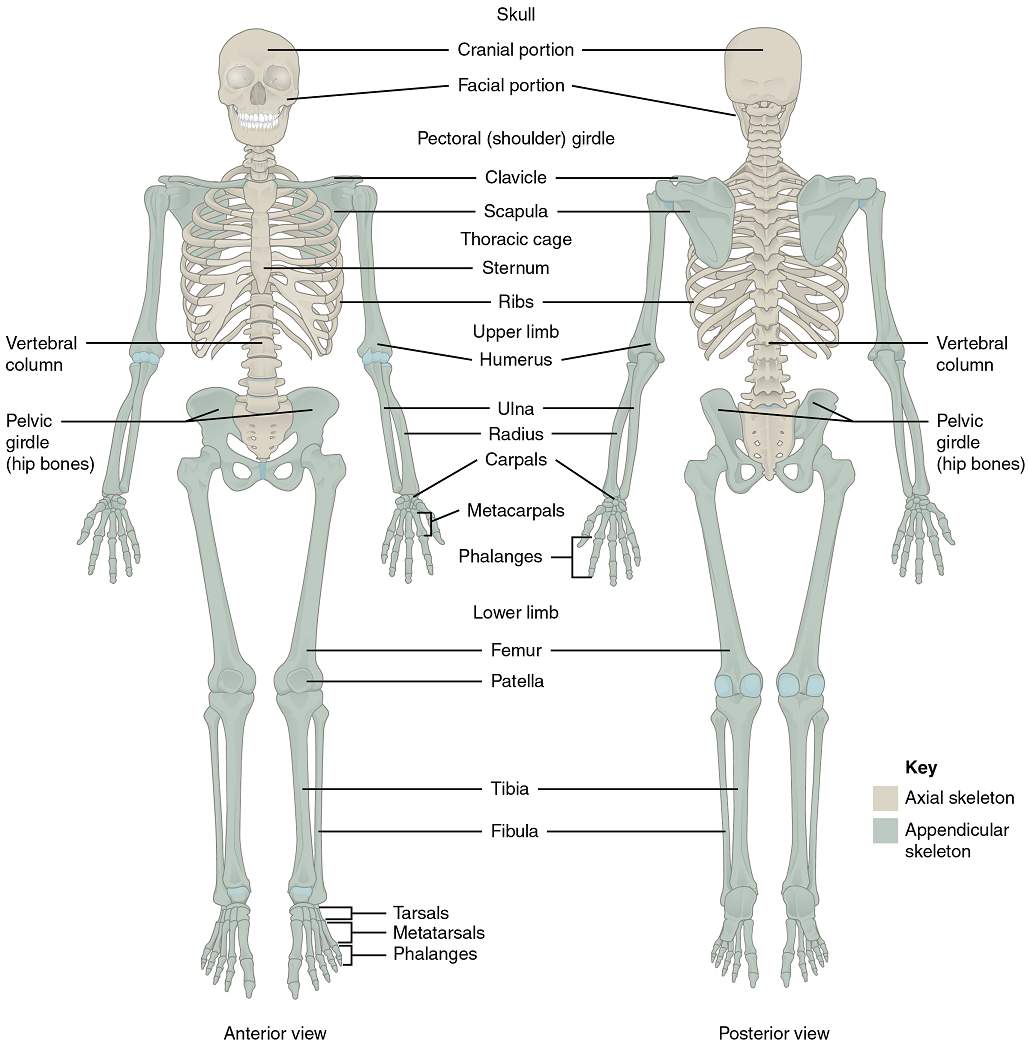
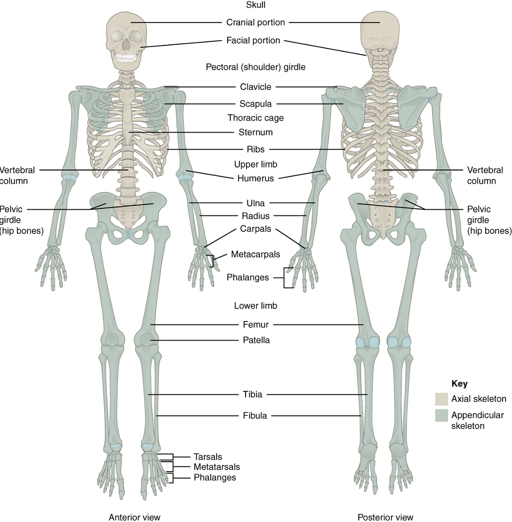
The skeletal system includes all of the bones, cartilages, and ligaments of the body that support and give shape to the body and body structures. The skeleton consists of the bones of the body. For adults, there are 206 bones in the skeleton. Younger individuals have higher numbers of bones because some bones fuse together during childhood and adolescence to form an adult bone. The skeleton is subdivided into two major divisions—the axial skeleton and the appendicular skeleton.The Axial Skeleton: The skeleton is subdivided into two major divisions—the axial skeleton and appendicular skeleton. The axial skeleton forms the vertical central axis of the body and includes all bones of the head, neck, chest, and back (Figure 10.17). It serves to protect the brain, spinal cord, heart, and lungs. It also serves as the attachment site for muscles that move the head, neck, and back, and for muscles that act across the shoulder and hip joints to move their corresponding limbs.The axial skeleton of the adult consists of 80 bones, including the skull, the vertebral column, and the thoracic cage. The skull is formed by 22 bones. Also associated with the head are an additional seven bones, including the hyoid bone and the ear ossicles (three small bones found in each middle ear). The vertebral column consists of 24 bones, each called a vertebra, plus the sacrum and coccyx. The thoracic cage includes the 12 pairs of ribs, and the sternum, the flattened bone of the anterior chest.The Appendicular Skeleton: The appendicular skeleton includes all bones of the upper and lower limbs, plus the bones that attach each limb to the axial skeleton (Figure 10.17). There are 126 bones in the appendicular skeleton of an adult. The bones of the appendicular skeleton are covered later in the unit.
Part 1: The Axial Skeleton
The Skull
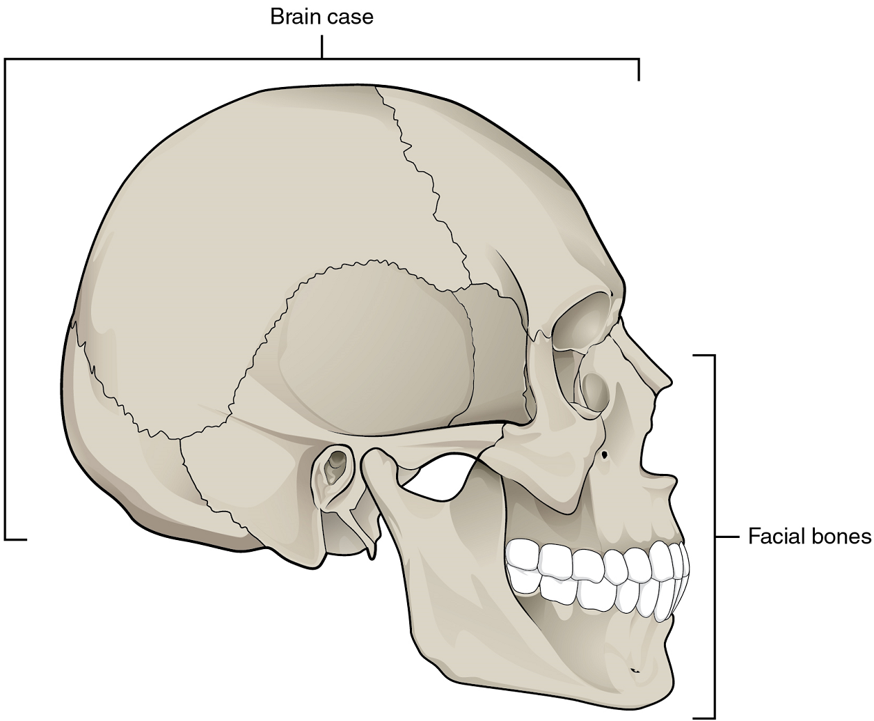
The cranium (skull) is the skeletal structure of the head that supports the face and protects the brain. It is subdivided into the facial bones and the braincase, or cranial vault (Figure 10.19). The facial bones underlie the facial structures, form the nasal cavity, enclose the eyeballs, and support the teeth of the upper and lower jaws. The rounded braincase surrounds and protects the brain and houses the middle and inner ear structures.
In the adult, the skull consists of 22 individual bones, 21 of which are immobile and united into a single unit. The 22nd bone is the mandible (lower jaw), which is the only moveable bone of the skull.
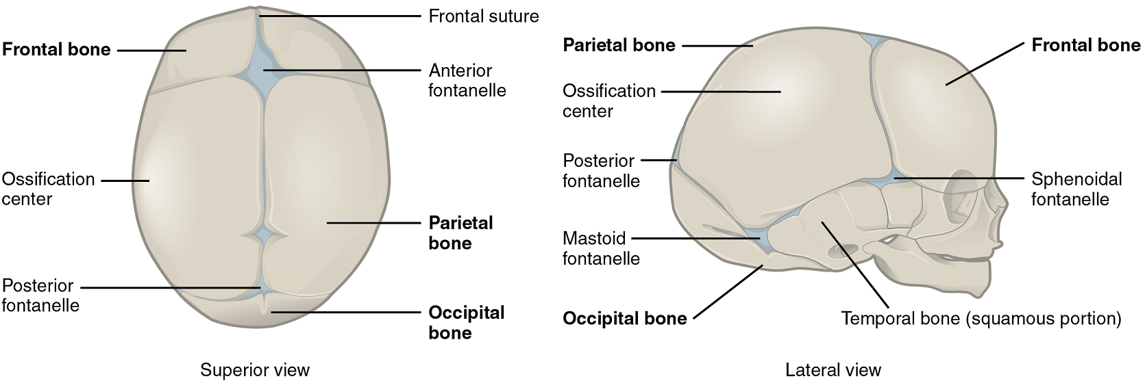
Development of the Skull: As the braincase bones grow in the fetal skull, they remain separated from each other by large areas of dense connective tissue, each of which is called a fontanelle (Figure 10.18). The fontanelles are the soft spots on an infant’s head. They are important during birth because these areas allow the skull to change shape as it squeezes through the birth canal. After birth, the fontanelles allow for continued growth and expansion of the skull as the brain enlarges. The largest fontanelle is located on the anterior head, at the junction of the frontal and parietal bones. The fontanelles decrease in size and disappear by age 2. However, the skull bones remained separated from each other at the sutures, which contain dense fibrous connective tissue that unites the adjacent bones. The connective tissue of the sutures allows for continued growth of the skull bones as the brain enlarges during childhood growth. This structure also means that, although the size of the cranium increases from birth to adulthood, proportionately, it does so less than other parts of the skeleton. The relative size of the cranium in proportion to the rest of the body therefore decreases with age from birth to adulthood.
Bones of the Braincase: The braincase contains and protects the brain (Figure 10.19). The interior space that is almost completely occupied by the brain is called the cranial cavity.
The braincase consists of eight bones (Figures 10.20 & 10.21). These include the paired parietal and temporal bones, plus the unpaired frontal, occipital, sphenoid, and ethmoid bones. For our purposes, we will not be specifying the details of the sphenoid and ethmoid bones.
1. Parietal Bone: The parietal bone forms most of the upper lateral side of the skull (Figures 10.21 & 10.22). These are paired bones, with the right and left parietal bones joining together at the top of the skull. Each parietal bone is also bounded anteriorly by the frontal bone, inferiorly by the temporal bone, and posteriorly by the occipital bone.
2. Temporal Bone: The temporal bone forms the lower lateral side of the skull (Figure 10.21). Common wisdom has it that the temporal bone (temporal = “time”) is so named because this area of the head (the temple) is where hair typically first turns gray, indicating the passage of time.
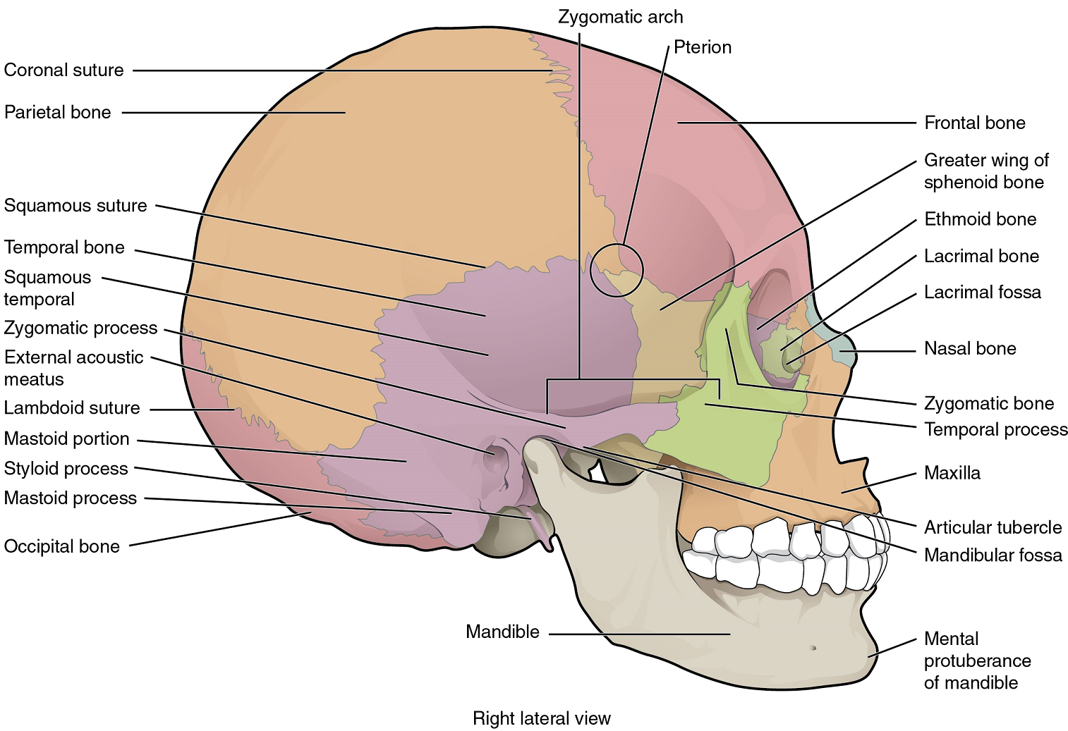
3. Frontal Bone: The frontal bone is the single bone that forms the forehead (Figure 10.20).
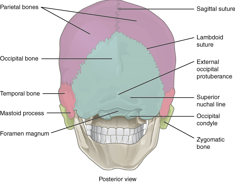
4. Occipital Bone: The occipital bone is the single bone that forms the posterior skull and posterior base of the cranial cavity (Figures 10.21 & 10.22). On its outside surface, at the posterior midline, is a small protrusion called the external occipital protuberance, which serves as an attachment site for a ligament of the posterior neck.
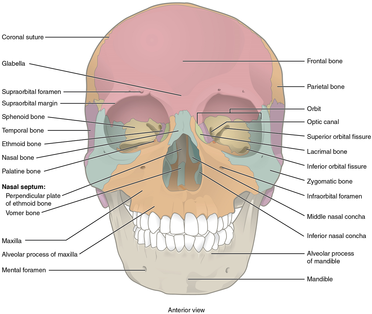
Facial Bones of the Skull: The facial bones of the skull form the upper and lower jaws, the nose, nasal cavity and nasal septum, and the orbit. The facial bones include 14 bones, with six paired bones and two unpaired bones (Figures 10.20 & 10.21). We will focus on the maxillary bones and the mandible bone.
1. Maxillary Bone: The maxillary bone, often referred to simply as the maxilla (plural = maxillae), is one of a pair that together form the upper jaw, much of the hard palate, the medial floor of the orbit, and the lateral base of the nose (Figures 10.20 & 10.21).
2. Mandible: The mandible forms the lower jaw and is the only moveable bone of the skull. At the time of birth, the mandible consists of paired right and left bones, but these fuse together during the first year to form the single U-shaped mandible of the adult skull (Figures 10.20 & 10.21).
The Bones of the Middle Ear: Three small bones (ossicles) are found on either side of the head in the middle ear. These are the malleus, incus, and stapes, and they function in transferring the vibrations from the eardrum (tympanic membrane) to the inner ear.
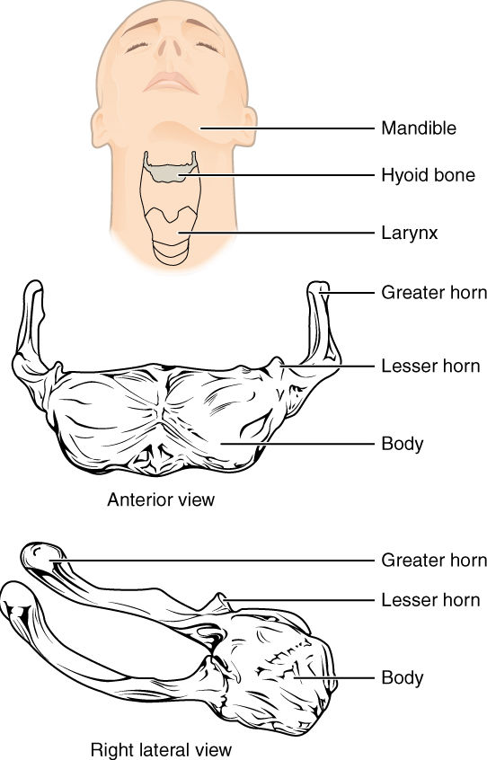
The Hyoid Bone: The hyoid bone is an independent bone that does not contact any other bone and thus is not part of the skull (Figure 10.23). It is a small U-shaped bone located in the upper neck near the level of the inferior mandible, with the tips of the “U” pointing posteriorly. The hyoid serves as the base for the tongue above and is attached to the larynx below and the pharynx posteriorly. The hyoid is held in position by a series of small muscles that attach to it either from above or below. These muscles act to move the hyoid up/down or forward/back. Movements of the hyoid are coordinated with movements of the tongue, larynx, and pharynx during swallowing and speaking.
The Vertebral Column
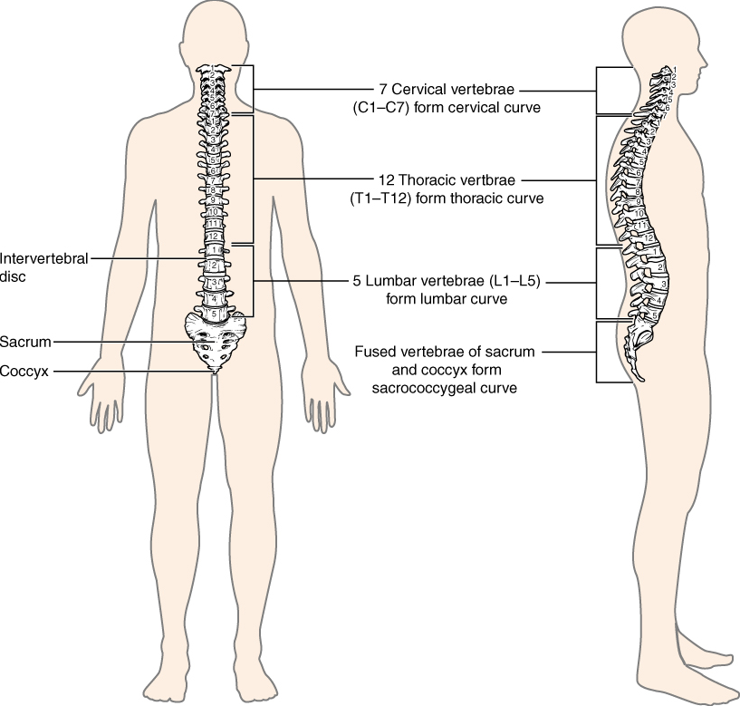
The vertebral column is also known as the spinal column or spine (Figure 10.24). It consists of a sequence of vertebrae (singular = vertebra), each of which is separated and united by an intervertebral disc. Together, the vertebrae and intervertebral discs form the vertebral column. It is a flexible column that supports the head, neck, and body and allows for their movements. It also protects the spinal cord, which passes down the back through openings in the vertebra.
Regions of the Vertebral Column: The vertebral column originally develops as a series of 33 vertebrae, but this number is eventually reduced to 24 vertebrae, plus the sacrum and coccyx. The vertebral column is subdivided into five regions, with the vertebrae in each area named for that region and numbered in descending order. In the neck, there are seven cervical vertebrae, each designated with the letter “C” followed by its number. Superiorly, the C1 vertebra articulates (forms a joint) with the occipital condyles of the skull. Inferiorly, C1 articulates with the C2 vertebra, and so on. Below these are the 12 thoracic vertebrae, designated T1–T12. The lower back contains the L1–L5 lumbar vertebrae. The single sacrum, which is also part of the pelvis, is formed by the fusion of five sacral vertebrae. Similarly, the coccyx, or tailbone, results from the fusion of four small coccygeal vertebrae. However, the sacral and coccygeal fusions do not start until age 20 and are not completed until middle age.
Curvatures of the Vertebral Column: The adult vertebral column does not form a straight line but instead has four curvatures along its length (see Figure 10.24). These curves increase the vertebral column’s strength, flexibility, and ability to absorb shock.
During fetal development, the body is flexed anteriorly into the fetal position, giving the entire vertebral column a single curvature that is concave anteriorly. In the adult, this fetal curvature is retained in two regions of the vertebral column as the thoracic curve, which involves the thoracic vertebrae, and the sacrococcygeal curve, formed by the sacrum and coccyx.
General Structure of a Vertebra: Within the different regions of the vertebral column, vertebrae vary in size and shape, but they all follow a similar structural pattern. A typical vertebra will consist of a body, a vertebral arch, and seven processes (Figure 10.25).
The body is the anterior portion of each vertebra and is the part that supports the body weight. Because of this, the vertebral bodies progressively increase in size and thickness going down the vertebral column. The bodies of adjacent vertebrae are separated and strongly united by an intervertebral disc.
The vertebral arch forms the posterior portion of each vertebra.
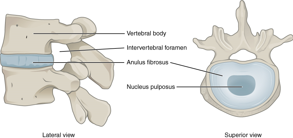
The large opening between the vertebral arch and body is the vertebral foramen, which contains the spinal cord. In the intact vertebral column, the vertebral foramina of all of the vertebrae align to form the vertebral (spinal) canal, which serves as the bony protection and passageway for the spinal cord down the back. When the vertebrae are aligned together in the vertebral column, notches in the margins of the pedicles of adjacent vertebrae together form an intervertebral foramen, the opening through which a spinal nerve exits from the vertebral column (Figure 10.26).
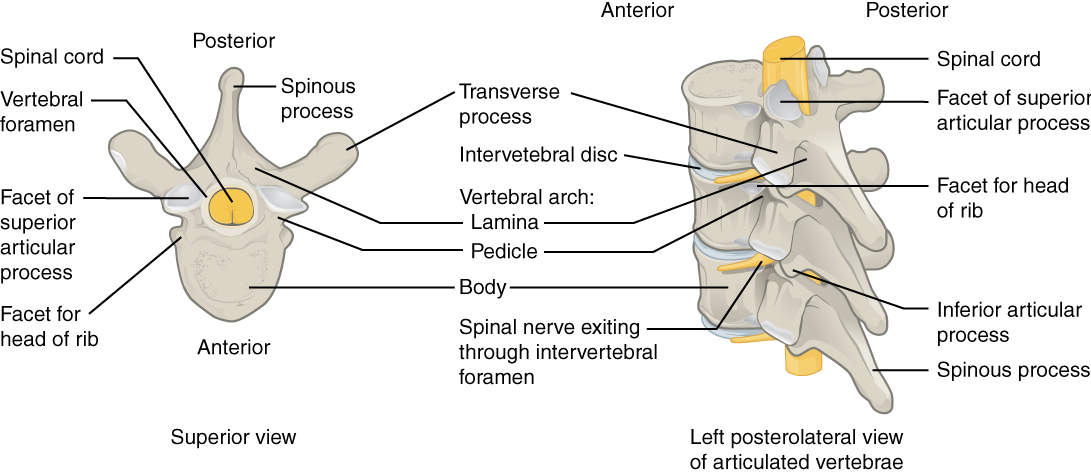
Seven processes arise from the vertebral arch. Each paired transverse process projects laterally and arises from the junction point between the pedicle and lamina. The single spinous process (vertebral spine) projects posteriorly at the midline of the back. The vertebral spines can easily be felt as a series of bumps just under the skin down the middle of the back. The transverse and spinous processes serve as important muscle attachment sites. A superior articular process extends or faces upward, and an inferior articular process faces or projects downward on each side of a vertebrae. The paired superior articular processes of one vertebra join with the corresponding paired inferior articular processes from the next higher vertebra. These junctions form slightly moveable joints between the adjacent vertebrae. The shape and orientation of the articular processes vary in different regions of the vertebral column and play a major role in determining the type and range of motion available in each region.
Regional Modifications of Vertebrae: In addition to the general characteristics of a typical vertebra described above, vertebrae also display characteristic size and structural features that vary between the different vertebral column regions. Thus, cervical vertebrae are smaller than lumbar vertebrae due to differences in the proportion of body weight that each will support. Thoracic vertebrae have sites for rib attachment, and the vertebrae that give rise to the sacrum and coccyx have fused together into single bones. We will focus on the anatomically distinct natures of the first two cervical vertebrae, the atlas and the axis.
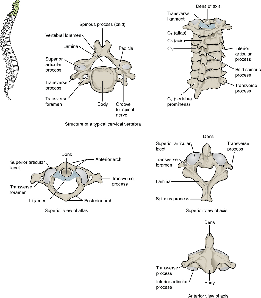
Cervical Vertebrae: Typical cervical vertebrae, such as C4 or C5, have several characteristic features that differentiate them from thoracic or lumbar vertebrae (Figure 10.27). Cervical vertebrae have a small body, reflecting the fact that they carry the least amount of body weight. Cervical vertebrae usually have a bifid (Y-shaped) spinous process. The transverse processes of the cervical vertebrae are sharply curved (U-shaped) to allow for passage of the cervical spinal nerves. Each transverse process also has an opening called the transverse foramen.
The first and second cervical vertebrae are further modified, giving each a distinctive appearance. The first cervical (C1) vertebra is also called the atlas, because this is the vertebra that supports the skull on top of the vertebral column (in Greek mythology, Atlas was the god who supported the heavens on his shoulders). The C1 vertebra does not have a body or spinous process. Instead, it is ring-shaped, consisting of an anterior arch and a posterior arch. The transverse processes of the atlas are longer and extend more laterally than do the transverse processes of any other cervical vertebrae. The superior articular processes face upward and are deeply curved for articulation with the occipital condyles on the base of the skull. The inferior articular processes are flat and face downward to join with the superior articular processes of the C2 vertebra.
The second cervical (C2) vertebra is called the axis, because it serves as the axis for rotation when turning the head right or left. The axis resembles typical cervical vertebrae in most respects but is easily distinguished by the dens (odontoid process), a bony projection that extends upward from the vertebral body. The dens joins with the inner aspect of the anterior arch of the atlas, where it is held in place by transverse ligament.
The Thoracic Cage
The thoracic cage (rib cage) forms the thorax (chest) portion of the body. It consists of the 12 pairs of ribs with their costal cartilages and the sternum (Figure 10.28). The ribs are anchored posteriorly to the 12 thoracic vertebrae (T1–T12). The thoracic cage protects the heart and lungs.
Sternum: The sternum is the elongated bony structure that anchors the anterior thoracic cage. It consists of three parts: the manubrium, body, and xiphoid process.
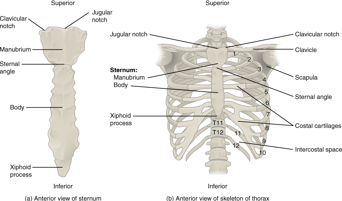
Ribs: Each rib is a curved, flattened bone that contributes to the wall of the thorax. The ribs articulate posteriorly with the T1–T12 thoracic vertebrae, and most attach anteriorly via their costal cartilages to the sternum. There are 12 pairs of ribs. The ribs are numbered 1–12 in accordance with the thoracic vertebrae.
The bony ribs do not extend anteriorly completely around to the sternum. Instead, each rib ends in a costal cartilage. These cartilages are made of hyaline cartilage and can extend for several inches. Most ribs are then attached, either directly or indirectly, to the sternum via their costal cartilage (Figure 10.28). The ribs are classified into three groups based on their relationship to the sternum.
Ribs 1–7 are classified as true ribs (vertebrosternal ribs). The costal cartilage from each of these ribs attaches directly to the sternum. Ribs 8–12 are called false ribs (vertebrochondral ribs). The costal cartilages from these ribs do not attach directly to the sternum. For ribs 8–10, the costal cartilages are attached to the cartilage of the next higher rib. Thus, the cartilage of rib 10 attaches to the cartilage of rib 9, rib 9 then attaches to rib 8, and rib 8 is attached to rib 7. The last two false ribs (11–12) are also called floating ribs (vertebral ribs). These are short ribs that do not attach to the sternum at all. Instead, their small costal cartilages terminate within the musculature of the lateral abdominal wall.
Test Your Knowledge
I. List all the components of the axial skeleton.
II. Specify the main functions of the axial skeleton. Explain how the overall shape of the axial skeleton is appropriate to its primary function.
III. Describe the structure and function of a typical vertebra and explain how these differ in the case of the atlas and axis.
Part 2. The Appendicular Skeleton
Attached to the axial skeleton are the limbs, whose 126 bones constitute the appendicular skeleton (Figure 10.29) These bones are divided into two groups: the bones that are located within the limbs themselves and the girdle bones that attach the limbs to the axial skeleton. The bones of the shoulder region form the pectoral girdle, which anchors the upper limb to the thoracic cage of the axial skeleton. The lower limb is attached to the vertebral column by the pelvic girdle.
Because of our upright stance, different functional demands are placed upon the upper and lower limbs. Thus, the bones of the lower limbs are adapted for weight-bearing support and stability, as well as for body locomotion via walking or running. In contrast, our upper limbs are not required for these functions. Instead, our upper limbs are highly mobile and can be utilized for a wide variety of activities. The large range of upper limb movements, coupled with the ability to easily manipulate objects with our hands and opposable thumbs, has allowed humans to construct the modern world in which we live.
The Pectoral Girdle
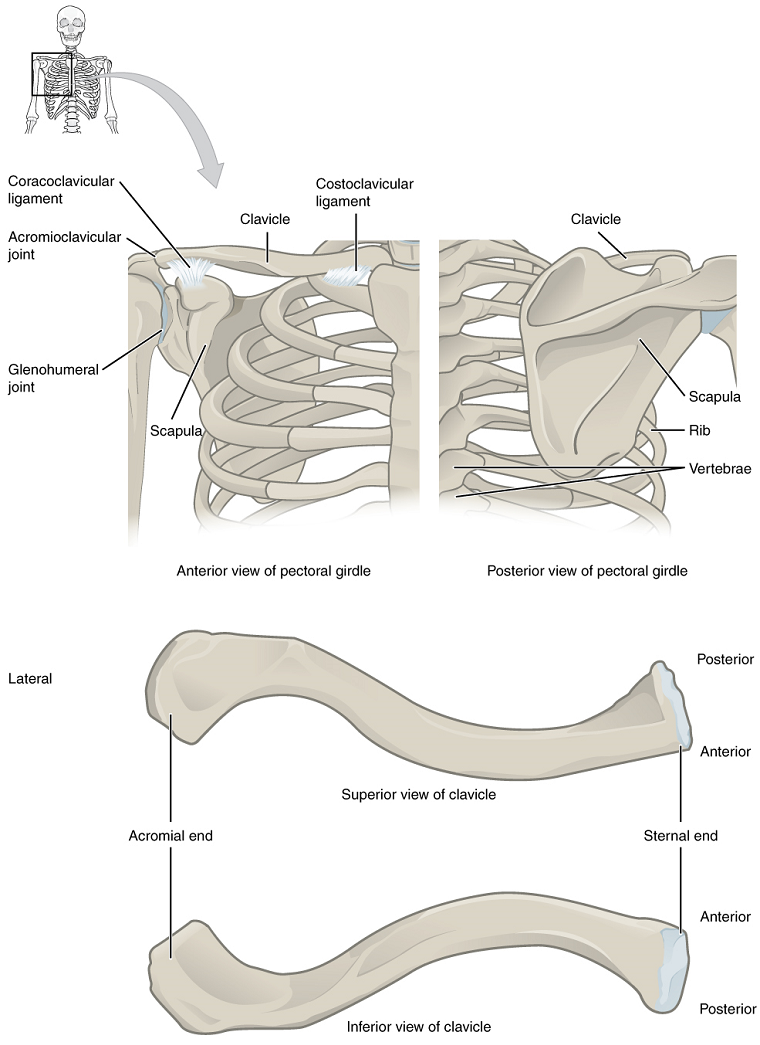
The bones that attach each upper limb to the axial skeleton form the pectoral girdle (shoulder girdle). This consists of two bones, the scapula and clavicle (Figure 10.30).
The clavicle (collarbone) is an S-shaped bone located on the anterior side of the shoulder. It is attached on its medial end to the sternum of the thoracic cage, which is part of the axial skeleton. The lateral end of the clavicle articulates (joins) with the scapula just above the shoulder joint. You can easily palpate, or feel with your fingers, the entire length of your clavicle.
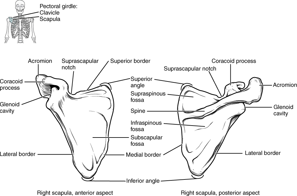
The scapula (shoulder blade) lies on the posterior aspect of the shoulder. It is supported by the clavicle, which also articulates with the humerus (upper arm bone) to form the shoulder joint. The scapula is a flat, triangular-shaped bone with a prominent ridge running across its posterior surface. This ridge extends out laterally, where it forms the bony tip of the shoulder and joins with the lateral end of the clavicle. By following along the clavicle, you can palpate out to the bony tip of the shoulder, and from there, you can move back across your posterior shoulder to follow the ridge of the scapula. Move your shoulder around and feel how the clavicle and scapula move together as a unit. Both of these bones serve as important attachment sites for muscles that aid with movements of the shoulder and arm (Figures 10.30 & 10.31).
The right and left pectoral girdles are not joined to each other, allowing each to operate independently. In addition, the clavicle of each pectoral girdle is anchored to the axial skeleton by a single, highly mobile joint. This allows for the extensive mobility of the entire pectoral girdle, which in turn enhances movements of the shoulder and upper limb.
Bones of the Upper Limb
The upper limb is divided into three regions. These consist of the arm, located between the shoulder and elbow joints; the forearm, which is between the elbow and wrist joints; and the hand, which is located distal to the wrist. There are 30 bones in each upper limb. The humerus is the single bone of the upper arm, and the ulna (medially) and the radius (laterally) are the paired bones of the forearm. The base of the hand contains eight bones, each called a carpal bone, and the palm of the hand is formed by five bones, each called a metacarpal bone. The fingers and thumb contain a total of 14 bones, each of which is a phalanx bone of the hand (Figure 10.29).
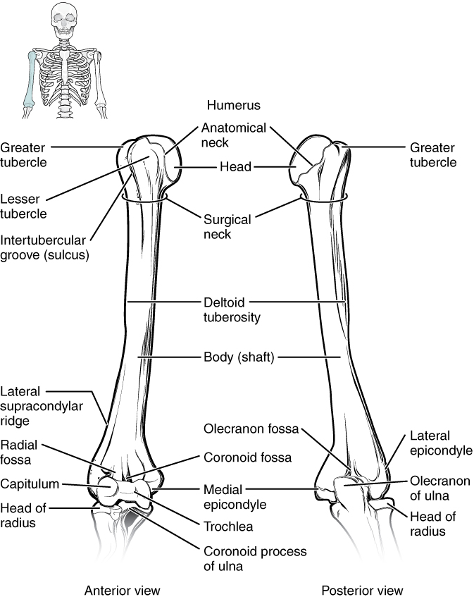
Humerus: The humerus is the single bone of the upper arm region (Figure 10.32). At its proximal end is the head of the humerus. This is the large, round, smooth region that faces medially. The head articulates with the glenoid cavity of the scapula to form the glenohumeral (shoulder) joint. Distally, the humerus becomes flattened and has two articulation areas, which join the ulna and radius bones of the forearm to form the elbow joint.
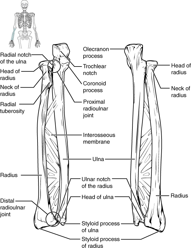
Ulna: The ulna is the medial bone of the forearm. It runs parallel to the radius, which is the lateral bone of the forearm (Figure 10.33). The proximal end of the ulna articulates with the humerus as part of the elbow joint.
Radius: The radius runs parallel to the ulna, on the lateral (thumb) side of the forearm (Figure 10.33). The head of the radius is a disc-shaped structure that forms the proximal end. The distal end of the radius has a smooth surface for articulation with two carpal bones to form the radiocarpal joint or wrist joint (Figures 10.34 & 10.35).
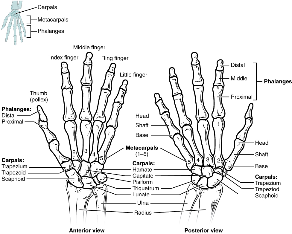
Carpal Bones: The wrist and base of the hand are formed by a series of eight small carpal bones (Figure 10.34). The carpal bones are arranged in two rows, forming a proximal row of four carpal bones and a distal row of four carpal bones.
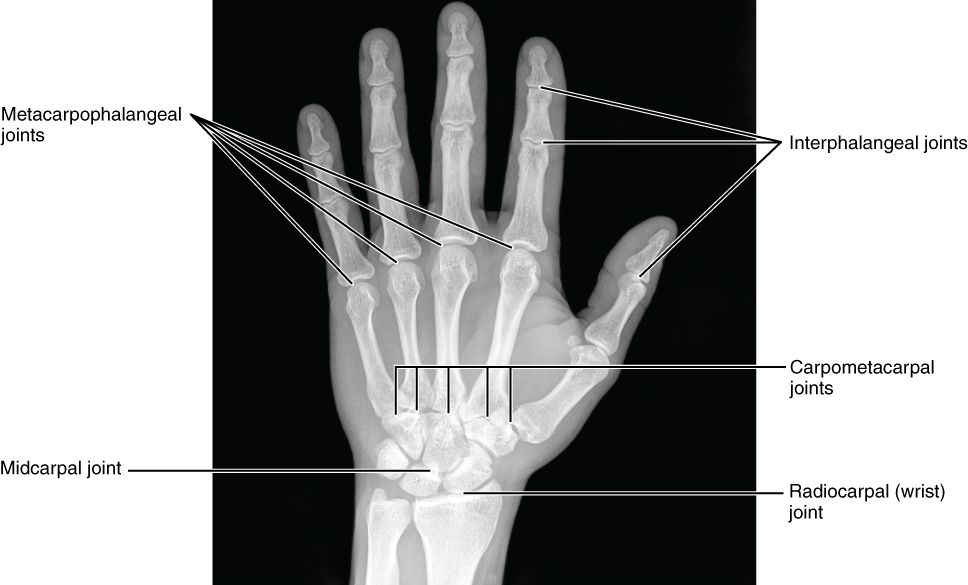
The carpal bones form the base of the hand. This can be seen in the radiograph (X-ray image) of the hand that shows the relationships of the hand bones to the skin creases of the hand (Figure 10.35).
Metacarpal Bones: The palm of the hand contains five elongated metacarpal bones. These bones lie between the carpal bones of the wrist and the bones of the fingers and thumb (Figure 10.34). The proximal end of each metacarpal bone articulates with one of the distal carpal bones. Each of these articulations is a carpometacarpal joint (Figure 10.35). The expanded distal end of each metacarpal bone articulates at the metacarpophalangeal joint with the proximal phalanx bone of the thumb or one of the fingers. The distal end also forms the knuckles of the hand, at the base of the fingers. The metacarpal bones are numbered 1–5, beginning at the thumb.
Phalanx Bones: The fingers and thumb contain 14 bones, each of which is called a phalanx bone (plural = phalanges), named after the ancient Greek phalanx (a rectangular block of soldiers). The thumb (pollex) is digit number 1 and has two phalanges, a proximal phalanx, and a distal phalanx bone (Figure 10.34). Digits 2 (index finger) through 5 (little finger) have three phalanges each, called the proximal, middle, and distal phalanx bones. An interphalangeal joint is one of the articulations between adjacent phalanges of the digits (Figure 10.35).
Test Your Knowledge
I. List all the components of the appendicular skeleton.
II. Specify the main functions of the appendicular skeleton. Explain how the overall shape of the appendicular skeleton is appropriate to its primary function.
Part 3: The Pelvic Girdle and Pelvis
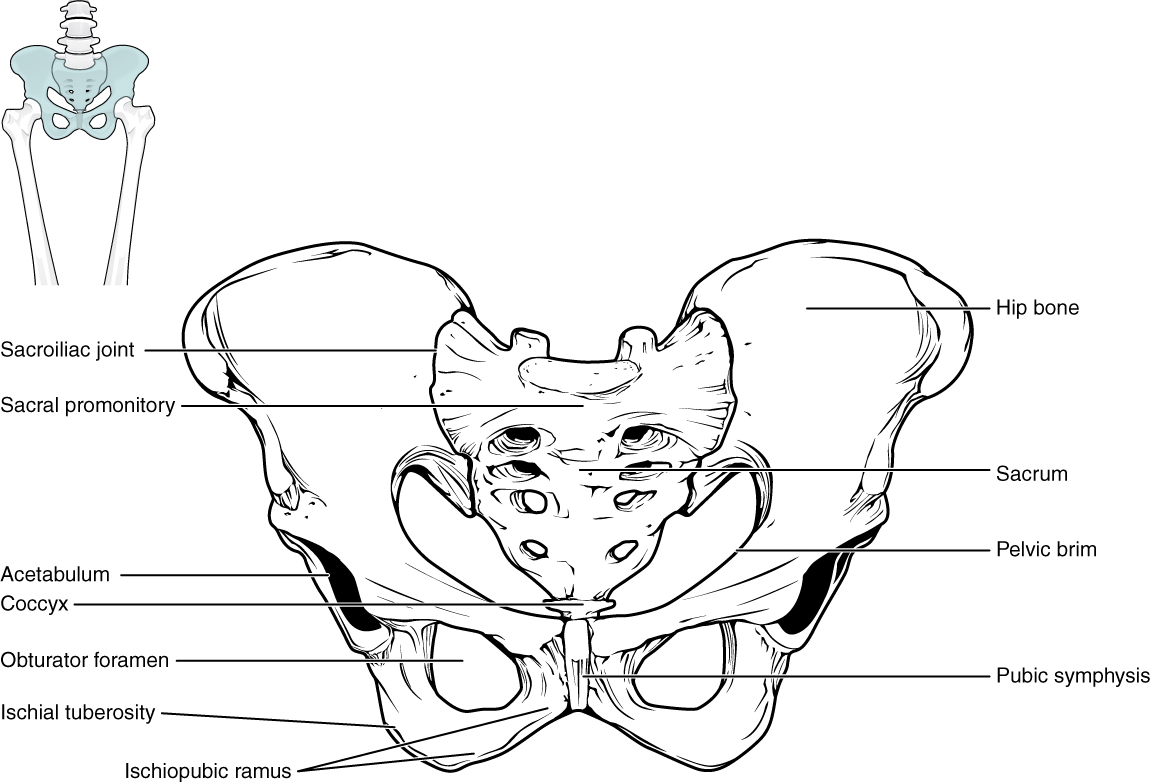
The pelvic girdle (hip girdle) is formed by a single bone, the hip bone or coxal bone (coxal = “hip”), which serves as the attachment point for each lower limb. Each hip bone, in turn, is firmly joined to the axial skeleton via its attachment to the sacrum of the vertebral column. The right and left hip bones also converge anteriorly to attach to each other. The bony pelvis is the entire structure formed by the two hip bones, the sacrum and the coccyx, which is attached inferiorly to the sacrum (Figure 10.36).
Unlike the bones of the pectoral girdle, which are highly mobile to enhance the range of upper limb movements, the bones of the pelvis are strongly united to each other to form a largely immobile, weight-bearing structure. This is important for stability because it enables the weight of the body to be easily transferred laterally from the vertebral column, through the pelvic girdle and hip joints, and into either lower limb whenever the other limb is not bearing weight. Thus, the immobility of the pelvis provides a strong foundation for the upper body as it rests on top of the mobile lower limbs.
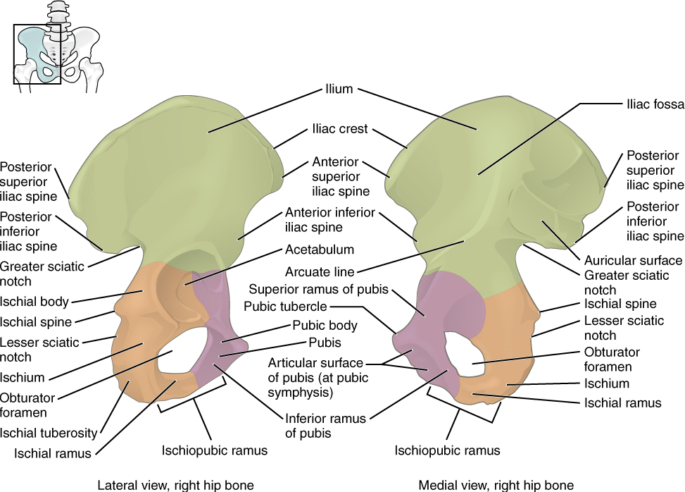
Hip Bone: The hip bone, or coxal bone, forms the pelvic girdle portion of the pelvis. The paired hip bones are the large, curved bones that form the lateral and anterior aspects of the pelvis. Each adult hip bone is formed by three separate bones that fuse together during the late teenage years. These bony components are the ilium, ischium, and pubis (Figure 10.37). These names are retained and used to define the three regions of the adult hip bone.
The ilium is the fan-like, superior region that forms the largest part of the hip bone. It is firmly united to the sacrum at the largely immobile sacroiliac joint (Figure 10.36). The ischium forms the posteroinferior region of each hip bone. It supports the body when sitting. The pubis forms the anterior portion of the hip bone. The pubis curves medially, where it joins to the pubis of the opposite hip bone at a specialized joint called the pubic symphysis.
Pelvis: The pelvis consists of four bones: the right and left hip bones, the sacrum, and the coccyx (Figure 10.36). The pelvis has several important functions. Its primary role is to support the weight of the upper body when sitting and to transfer this weight to the lower limbs when standing. It serves as an attachment point for trunk and lower limb muscles and also protects the internal pelvic organs.
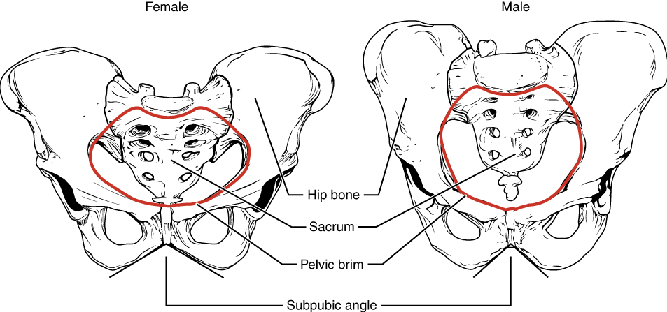
Comparison of the Female and Male Pelvis: The differences between the adult female and male pelvis relate to function and body size. In general, the bones of the male pelvis are thicker and heavier, adapted for support of the male’s heavier physical build and stronger muscles; this average size difference is generally true of other bones of the skeleton as well. The pelvis does show more robust differences between males and females due to its functional relationship to bipedal movement (requiring a relatively narrow pelvis) and birth of infants with large brains (requiring a relatively broad pelvis). Because the female pelvis is adapted for childbirth, it is wider than the male pelvis, as evidenced by the distance between the anterior superior iliac spines (Figure 10.38). The ischial tuberosities of females are also farther apart, which increases the size of the pelvic outlet. Because of this increased pelvic width, the subpubic angle is larger in females (greater than 80 degrees) than it is in males (less than 70 degrees). The female sacrum is wider, shorter, and less curved, and the sacral promontory projects less into the pelvic cavity, thus giving the female pelvic inlet (pelvic brim) a more rounded or oval shape compared to males. The pelvic cavity of females is also wider and shallower than the narrower, deeper, and tapering lesser pelvis of males. The greater sciatic notch of the male hip bone is narrower and deeper than the broader notch of females. Because of the obvious differences between female and male hip bones, this is the one bone of the body that allows for the most accurate sex determination. Table 10.4 provides an overview of the general differences between the female and male pelvis.
| Female pelvis | Male pelvis | |
|---|---|---|
| Pelvic weight | Bones are lighter and thinner | Bones are thicker and heavier |
| Pelvis inlet shape | Round or oval | Heart-shaped |
| Lesser pelvic cavity shape | Shorter and wider | Longer and narrower |
| Subpubic angle | Greater than 80 degrees | Less than 70 degrees |
| Pelvic outlet shape | Rounded and larger | Smaller |
Test Your Knowledge
I. Describe the differences between an average male pelvis and an average female pelvis.
II. Why are male and female pelves generally shaped differently? For each sex difference noted in the question above, briefly explain the functional relevance of that sex difference specifically.
Part 4: Bones of the Lower Limb
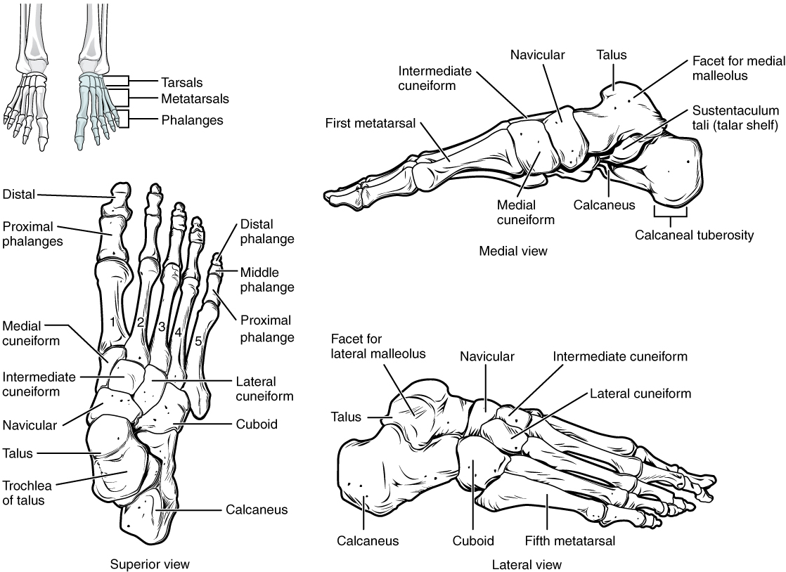
Like the upper limb, the lower limb is divided into three regions. The thigh is the portion of the lower limb located between the hip joint and knee joint. The leg is specifically the region between the knee joint and the ankle joint. Distal to the ankle is the foot. The lower limb contains 30 bones. These are the femur, patella, tibia, fibula, tarsal bones, metatarsal bones, and phalanges (Figure 10.29). The femur is the single bone of the thigh. The patella is the kneecap and articulates with the distal femur. The tibia is the larger, weight-bearing bone located on the medial side of the leg, and the fibula is the thin bone of the lateral leg. The bones of the foot are divided into three groups. The posterior portion of the foot is formed by a group of seven bones, each of which is known as a tarsal bone, whereas the mid-foot contains five elongated bones, each of which is a metatarsal bone. The toes contain 14 small bones, each of which is a phalanx bone of the foot.
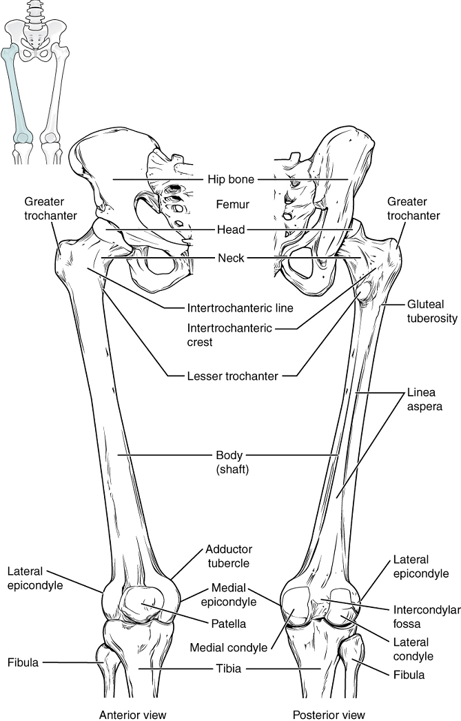
Femur: The femur, or thigh bone, is the single bone of the thigh region (Figure 10.39). It is the longest and strongest bone of the body and accounts for approximately one-quarter of a person’s total height. The rounded, proximal end is the head of the femur, which articulates with the acetabulum of the hip bone to form the hip joint.
Patella: The patella (kneecap) is the largest sesamoid bone of the body (see Figure 10.39). A sesamoid bone is a bone that is incorporated into the tendon of a muscle where that tendon crosses a joint. The sesamoid bone articulates with the underlying bones to prevent damage to the muscle tendon due to rubbing against the bones during movements of the joint. The patella is found in the tendon of the quadriceps femoris muscle, the large muscle of the anterior thigh that passes across the anterior knee to attach to the tibia. The patella articulates with the patellar surface of the femur and thus prevents rubbing of the muscle tendon against the distal femur. The patella also lifts the tendon away from the knee joint, which increases the leverage power of the quadriceps femoris muscle as it acts across the knee. The patella does not articulate with the tibia.
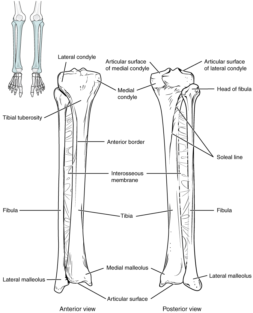
Tibia: The tibia (shin bone) is the medial bone of the leg and is larger than the fibula, with which it is paired (Figure 10.40). The tibia is the main weight-bearing bone of the lower leg and the second longest bone of the body, after the femur. The medial side of the tibia is located immediately under the skin, allowing it to be easily palpated down the entire length of the medial leg.
Fibula: The fibula is the slender bone located on the lateral side of the leg (Figure 10.40). The fibula does not bear weight. It serves primarily for muscle attachments and thus is largely surrounded by muscles. Only the proximal and distal ends of the fibula can be palpated.
Tarsal Bones: The posterior half of the foot is formed by seven tarsal bones (Figure 10.43). The most superior tarsal bone, the talus, articulates with the tibia and fibula to form the ankle joint. Inferiorly, the talus articulates with the calcaneus (heel bone), the largest bone of the foot, which forms the heel. Body weight is transferred from the tibia to the talus to the calcaneus, which rests on the ground.
Metatarsal Bones: The anterior half of the foot is formed by the five metatarsal bones, which are located between the tarsal bones of the posterior foot and the phalanges of the toes (Figure 10.41). These elongated bones are numbered 1–5, starting with the medial side of the foot.
Phalanx bones: The toes contain a total of 14 phalanx bones (phalanges), arranged in a similar manner as the phalanges of the fingers (Figure 10.41). The toes are numbered 1–5, starting with the big toe (hallux). The big toe has two phalanx bones, the proximal and distal phalanges. The remaining toes all have proximal, middle, and distal phalanges. A joint between adjacent phalanx bones is called an interphalangeal joint.
Test Your Knowledge
I. Identify and describe the bones of the lower limbs.
II. Identify the three groups of the foot bones.
Practice
For the activity below, drag the structure names to the correct empty boxes on the figure.
Image Descriptions

Figure 10.4 image description: There are different types of bone classifications: Flat bone, an example is the sternum. Long bone, an example is the femur.  Sesamoid bone, an example is the patella, also the Lateral cuneiform, intermediate cuneiform, and medial cuneiform. Irregular bone, an example is a vertebra. Short bone, an example is a tarsal. [Return to image.]

Figure 10.5 image description:  The anatomy of a long bone has many components. The proximal epiphysis contains the articular cartilage. The metaphysis is the neck of the long bone between the proximal and distal epiphysis and the diaphysis; it contains the growth plate. The diaphysis is the central shaft-like portion of the bone, made up of compact bone forming the hollow center called the medullary cavity. The medullary cavity is lined with a thin epithelial membrane that is filled; this cavity is filled with blood cell–producing red bone marrow in children, and in adults, most of this marrow has turned to yellow marrow. [Return to image.]

Figure 10.8 image description: Osteogenic cells are undifferentiated and develop into osteoblasts. When osteoblasts get trapped within the calcified matrix, their structure and function changes, and they become osteocytes. Osteoclasts develop from monocytes and macrophages and differ in appearance from other bone cells. [Return to image.]

Figure 10.12 image description: (a) Mesenchymal cells group into clusters, and ossification centers form. (b) Secreted osteoid traps osteoblasts, which then become osteocytes. (c) Trabecular matrix and periosteum form. (d) Compact bone develops superficial to the trabecular bone, and crowded blood vessels condense into red marrow. [Return to image.]

Figure 10.13 image description: (a) Mesenchymal cells differentiate into chondroblasts that secrete cartilage matrix to form a hyaline cartilage model of the future bony skeleton. The perichondrium forms. (b) The cartilage model starts to calcify; chondrocytes begin to die, and the cartilage begins to degenerate. (c) Periosteal bud penetrates cartilage of the diaphysis; the primary ossification center develops. A bony collar develops around the diaphysis of the bone. Perichondrium transforms into periosteum. (d) Cartilage continues to grow at the ends of the bone. Medullary cavity forms. (e) Periosteal buds penetrate cartilage at epiphyses; secondary ossification centers develop. (f) Cartilage remains at the epiphyseal (growth) plate and at the joint surface as articular cartilage. [Return to image.]

Figure 10.15 image description: (a) Epiphyseal plates are visible in a growing bone. (b) Epiphyseal lines are the remnants of epiphyseal plates in a mature bone. [Return to image.]

Figure 10.16 image description: (a) Closed fracture, (b) open fracture, (c) transverse fracture, (d) spiral fracture, (e) comminuted fracture, (f) impacted fracture, (g) greenstick fracture, and (h) oblique fracture. [Return to image.]

Figure 10.17 image description: The axial skeleton consists of the skull, vertebral column (including the sacrum and coccyx), and the thoracic cage, formed by the ribs and sternum. The appendicular skeleton is made up of all bones of the upper and lower limbs. [Return to image.]
Figure 10.18 image description: Large areas that are filled with fibrous connective tissue. The fontanelles allow for continued growth of the skull after birth. At the time of birth, the facial bones are small and underdeveloped, and the mastoid process has not yet formed. [Return to image.]
Figure 10.21 image description: The zygomatic arch is formed jointly by the zygomatic process of the temporal bone and the temporal process of the zygomatic bone. The shallow space above the zygomatic arch is the temporal fossa. The space inferior to the zygomatic arch and deep to the posterior mandible is the infratemporal fossa. [Return to image.]
Figure 10.22 image description: The posterior view if the skull includes the parietal bone, occipital bone, temporal bone, mastoid process, foramen magnum, zygomatic bone, superior nuchal line, external occipital protuberance, lambdoid suture, and sagittal suture. [Return to image.]

Figure 10.23 image description: The hyoid bone provides attachments for muscles that act on the tongue, larynx, and pharynx. [Return to image.]
Figure 10.24 image description: The vertebrae are divided into three regions: cervical C1–C7 vertebrae, thoracic T1–T12 vertebrae, and lumbar L1–L5 vertebrae. The vertebral column is curved, with two primary curvatures (thoracic and sacrococcygeal curves) and two secondary curvatures (cervical and lumbar curves). [Return to image.]
Figure 10.25 image description: The arch is formed by the paired pedicles and paired laminae. Arising from the vertebral arch are the transverse, spinous, superior articular, and inferior articular processes. The vertebral foramen provides for passage of the spinal cord. Each spinal nerve exits through an intervertebral foramen, located between adjacent vertebrae. Intervertebral discs unite the bodies of adjacent vertebrae. [Return to image.]
Figure 10.26 image description: The intervertebral disc provides padding and allows for movements between adjacent vertebrae. The disc consists of a fibrous outer layer called the annulus fibrosus and a gel-like center called the nucleus pulposus. The intervertebral foramen is the opening formed between adjacent vertebrae for the exit of a spinal nerve. [Return to image.]
Figure 10.27 image description: The atlas (C1 vertebra) does not have a body or spinous process. It consists of an anterior and a posterior arch and elongated transverse processes. The axis (C2 vertebra) has the upward projecting dens, which articulates with the anterior arch of the atlas. [Return to image.]
Figure 10.28 image description: The ribs are anchored posteriorly to the 12 thoracic vertebrae. The sternum consists of the manubrium, body, and xiphoid process. The ribs are classified as true ribs (1–7) and false ribs (8–12). The last two pairs of false ribs are also known as floating ribs (11–12). [Return to image.]
Figure 10.41 image description: The posterior foot is formed by the seven tarsal bones. The mid-foot has the five metatarsal bones. The toes contain the phalanges. [Return to image.]
In neurons, that portion of the cell that contains the nucleus; the cell body, as opposed to the cell processes (axons and dendrites).
Regions of the nervous system containing cell bodies of neurons with few or no myelinated axons; actually may be more pink or tan in color but called gray in contrast to white matter.
Regions of the nervous system containing cell bodies of neurons with few or no myelinated axons; actually may be more pink or tan in color but called gray in contrast to white matter.
In cells, an extension of a cell body; in the case of neurons, this includes the axon and dendrites.
Three small bones in the middle ear.
Regions of the nervous system containing mostly myelinated axons, making the tissue appear white because of the high lipid content of myelin.
Regions of the nervous system containing mostly myelinated axons, making the tissue appear white because of the high lipid content of myelin.
Lowest part of the vertebral column; 'tailbone'
Left or right central abdominopelvic region.
Describes the front or direction toward the front of the body; also referred to as ventral.
(In nervous system) a localized collection of neuron cell bodies that are functionally related; a “center” of neural function (plural= nuclei).
(In nervous system) a localized collection of neuron cell bodies that are functionally related; a “center” of neural function (plural= nuclei).
Localized collection of neuron cell bodies in the peripheral nervous system.
Localized collection of neuron cell bodies in the peripheral nervous system.
Division of the posterior (dorsal) cavity that houses the brain.
Cord-like bundle of axons located in the peripheral nervous system that transmits sensory input and response output to and from the central nervous system.
Cord-like bundle of axons located in the peripheral nervous system that transmits sensory input and response output to and from the central nervous system.
Cord-like bundle of axons located in the peripheral nervous system that transmits sensory input and response output to and from the central nervous system.
Cord-like bundle of axons located in the peripheral nervous system that transmits sensory input and response output to and from the central nervous system.
Functional division of the nervous system that is concerned with conscious perception, voluntary movement, and skeletal muscle reflexes.
Functional division of the nervous system that is concerned with conscious perception, voluntary movement, and skeletal muscle reflexes.
Paired bones that form the lateral, inferior portions of the skull, with squamous, mastoid, and petrous portions.
Functional division of the nervous system that is responsible for homeostatic reflexes that coordinate control of cardiac and smooth muscle, as well as glandular tissue.
Functional division of the nervous system that is responsible for homeostatic reflexes that coordinate control of cardiac and smooth muscle, as well as glandular tissue.
Tapering of the neuron cell body that gives rise to the axon.
Information flow in one direction.
Tapering of the neuron cell body that gives rise to the axon.
Information flow in one direction.
General anatomical term for a hole or opening (usually in bone. Plural = foramina).
Glial cell type in the PNS that provides the myelin insulation for axons in nerves.
Single stretch of the axon insulated by myelin and bounded by nodes of Ranvier at either end (except for the first, which is after the initial segment, and the last, which is followed by the axon terminal).
Single stretch of the axon insulated by myelin and bounded by nodes of Ranvier at either end (except for the first, which is after the initial segment, and the last, which is followed by the axon terminal).
End of the axon, where there are usually several branches extending toward the target cell.
End of the axon, where there are usually several branches extending toward the target cell.
Swelling at the end of an axon where neurotransmitter molecules are released onto a target cell across a synapse.
Swelling at the end of an axon where neurotransmitter molecules are released onto a target cell across a synapse.
Shape of a neuron that has multiple processes—the axon and two or more dendrites.
Shape of a neuron that has multiple processes—the axon and two or more dendrites.
Region of the adult brain connected primarily to the pons that developed from the metencephalon (along with the pons) and is largely responsible for comparing information from the cerebrum with sensory feedback from the periphery through the spinal cord.
Region of the adult brain connected primarily to the pons that developed from the metencephalon (along with the pons) and is largely responsible for comparing information from the cerebrum with sensory feedback from the periphery through the spinal cord.
Glial cell type in the CNS that provides the myelin insulation for axons in tracts.
Glial cell type in the CNS that provides the myelin insulation for axons in tracts.
Glial cell type in the PNS that provides the myelin insulation for axons in nerves.
Lipid-rich layer of insulation that surrounds an axon, formed by oligodendrocytes in the CNS and Schwann cells in the PNS; facilitates the transmission of electrical signals.
Lipid-rich layer of insulation that surrounds an axon, formed by oligodendrocytes in the CNS and Schwann cells in the PNS; facilitates the transmission of electrical signals.
Region of the adult brain that develops from the telencephalon and is responsible for higher neurological functions such as memory, emotion, and consciousness.
Region of the adult brain that develops from the telencephalon and is responsible for higher neurological functions such as memory, emotion, and consciousness.
Homeostatic mechanism that tends to stabilize an upset in the body’s physiological condition by preventing an excessive response to a stimulus, typically as the stimulus is removed.
Describes the middle or direction toward the middle of the body.
Central abdominopelvic region including the belly button.
See parasagittal plane
Describes the back or direction toward the back of the body; also referred to as dorsal.
Describes the side or direction toward the side of the body.
See frontal plane
See parasagittal plane
Abdominopelvic region located in the central superior area below the xiphoid process.
See frontal plane
One half of the bilaterally symmetrical cerebrum.
One half of the bilaterally symmetrical cerebrum.
Describes a position in a limb that is nearer to the point of attachment or the trunk of the body.
Outer gray matter covering the forebrain, marked by wrinkles and folds known as gyri and sulci.
Describes a position in a limb that is farther from the point of attachment or the trunk of the body.
Abdominopelvic region located in the central superior area below the xiphoid process.
Thumb
Outer gray matter covering the forebrain, marked by wrinkles and folds known as gyri and sulci.
Ridge formed by convolutions on the surface of the cerebrum or cerebellum.
Ridge formed by convolutions on the surface of the cerebrum or cerebellum.
Groove formed by convolutions in the surface of the cerebral cortex.
Groove formed by convolutions in the surface of the cerebral cortex.
Nuclei of the cerebrum (with a few components in the upper brain stem and diencephalon) that are responsible for assessing cortical movement commands and comparing them with the general state of the individual through broad modulatory activity of dopamine neurons; largely related to motor functions, as evidenced through the symptoms of Parkinson’s and Huntington’s diseases.
Nuclei of the cerebrum (with a few components in the upper brain stem and diencephalon) that are responsible for assessing cortical movement commands and comparing them with the general state of the individual through broad modulatory activity of dopamine neurons; largely related to motor functions, as evidenced through the symptoms of Parkinson’s and Huntington’s diseases.
An important neurotransmitter.
Thigh bone; the single bone of the thigh.
Knee cap.
Abdominopelvic region (left or right) located under the lowest ribs in the superior corners of the abdominopelvic cavity.
Abdominopelvic region (left or right) located under the lowest ribs in the superior corners of the abdominopelvic cavity.
Left or right central abdominopelvic region.
Lowest part of the vertebral column; 'tailbone'
An important neurotransmitter.
Region of the adult brain that retains its name from embryonic development and includes the thalamus and hypothalamus.
Region of the adult brain that retains its name from embryonic development and includes the thalamus and hypothalamus.
Referring to the sense of smell.
Big toe
