Functions of the Respiratory System
A typical human cannot survive without breathing for more than 3 minutes, and even if you wanted to hold your breath longer, your autonomic nervous system would take control. This is because cells need to maintain oxidative metabolism for energy production that continuously regenerates adenosine triphosphate (ATP). For oxidative phosphorylation to occur, oxygen is used as a reactant and carbon dioxide is released as a waste product. You may be surprised to learn that although oxygen is a critical need for cells, it is actually the accumulation of carbon dioxide that primarily drives your need to breathe. Carbon dioxide is exhaled and oxygen is inhaled through the respiratory system, which includes muscles to move air into and out of the lungs, passageways through which air moves, and microscopic gas exchange surfaces covered by capillaries. The cardiovascular system transports gases from the lungs to tissues throughout the body and vice versa. A variety of diseases can affect the respiratory system, such as asthma, emphysema, chronic obstructive pulmonary disorder (COPD), and lung cancer. All of these conditions affect the gas exchange process and result in labored breathing and other difficulties.
The major organs of the respiratory system function primarily to provide oxygen to body tissues for cellular respiration, remove the waste product carbon dioxide, and help to maintain acid-base balance. Portions of the respiratory system are also used for non-vital functions, such as sensing odors, producing speech, and for straining, such as during childbirth or coughing.
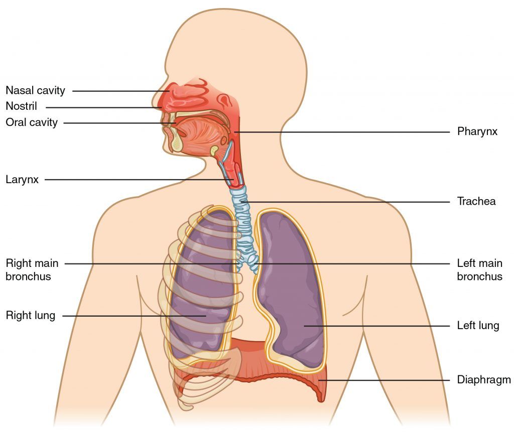
Major organs of the respiratory system (Source: University of Hawaii @ Manoa, CC-BY-NC-SA)
The major respiratory structures span the nasal cavity to the diaphragm. Functionally, the respiratory system can be divided into a conducting zone and a respiratory zone. The conducting zone of the respiratory system includes the organs and structures not directly involved in gas exchange (trachea and bronchi). The gas exchange occurs in the respiratory zone.
Conducting Zone
The major functions of the conducting zone are to provide a route for incoming and outgoing air, remove debris and pathogens from the incoming air, and warm and humidify the incoming air. Several structures within the conducting zone perform other functions as well. The epithelium of the nasal passages, for example, is essential to sensing odors, and the bronchial epithelium that lines the lungs can metabolize some airborne carcinogens. The conducting zone includes the nose and its adjacent structures, the pharynx, the larynx, the trachea, and the bronchi.
Respiratory Zone
In contrast to the conducting zone, the respiratory zone includes structures that are directly involved in gas exchange. The respiratory zone begins where the terminal bronchioles join a respiratory bronchiole, the smallest type of bronchiole (Figure 2.15 “Respiratory Zone”), which then leads to an alveolar duct, opening into a cluster of alveoli.
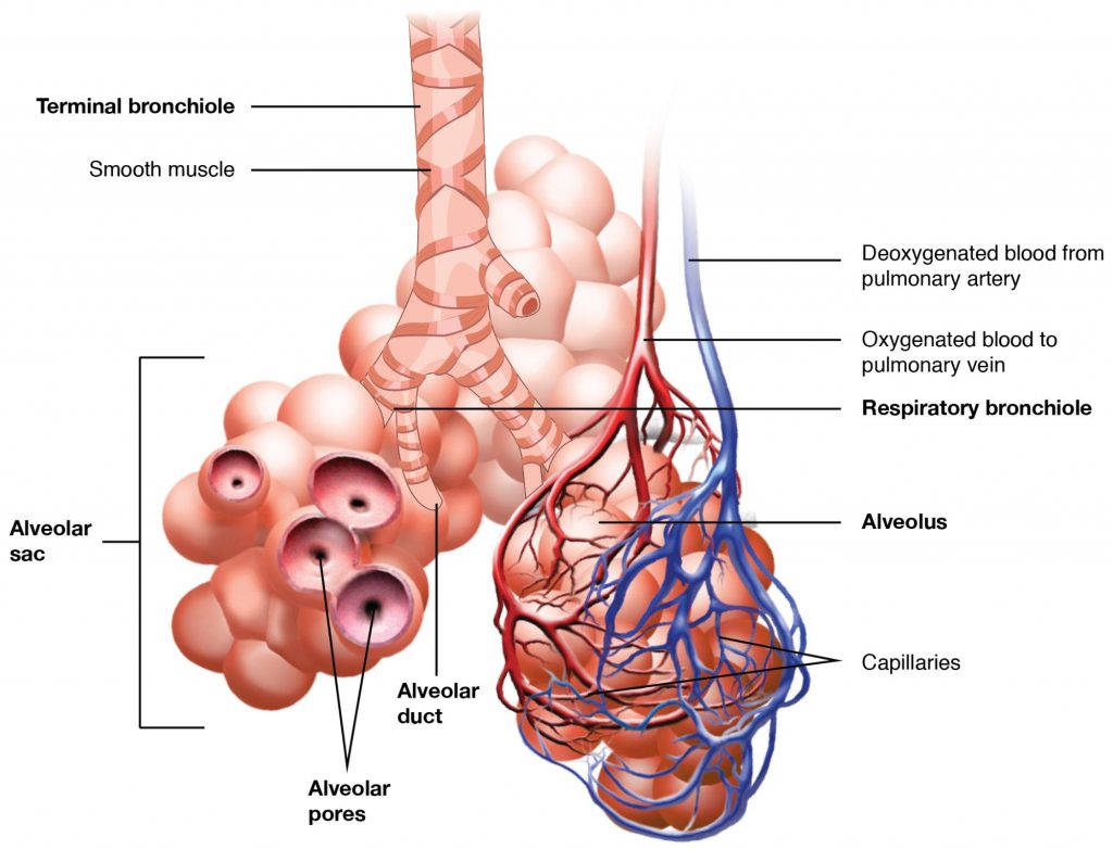
Bronchioles lead to alveolar sacs in the respiratory zone, where gas exchange occurs. (Source: University of Hawaii @ Manoa, CC-BY-NC-SA)
Alveoli
An alveolar duct is a tube composed of smooth muscle and connective tissue, which opens into a cluster of alveoli. An alveolus is one of the many small, grape-like sacs that are attached to the alveolar ducts.
An alveolar sac is a cluster of many individual alveoli that are responsible for gas exchange. An alveolus is approximately 200 μm in diameter with elastic walls that allow the alveolus to stretch during air intake, which greatly increases the surface area available for gas exchange. Alveoli are connected to their neighbors by alveolar pores, which help maintain equal air pressure throughout the alveoli and lungs.
(Source: Wikimedia Commons, U.S. National Institute of Health, Public Domain)
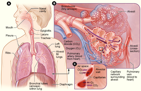
Figure 2.16 shows the location of the respiratory structures in the body. Figure B is an enlarged view of the airways, alveoli (air sacs), and capillaries (tiny blood vessels). Figure C is a close-up view of gas exchange between the capillaries and alveoli. CO2 is carbon dioxide, and O2 is oxygen.
A major organ of the respiratory system, each lung houses structures of both the conducting and respiratory zones. The lungs’ main function is to exchange oxygen and carbon dioxide with air from the atmosphere. To this end, the lungs exchange respiratory gases across a very large epithelial surface area—about 70 square meters—that is highly permeable to gases.
Gross Anatomy of the Lungs
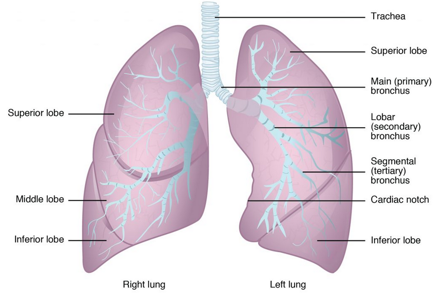
The lungs are paired, pyramid-shaped organs that are connected to the trachea by the right and left bronchi; below the lungs is the diaphragm, a flat, dome-shaped muscle located at the base of the lungs and thoracic cavity.
Bronchioles lead to alveolar sacs in the respiratory zone, where gas exchange occurs. (Source: University of Hawaii @ Manoa, CC-BY-NC-SA)
Each lung is composed of smaller units called lobes. Fissures separate these lobes from each other. The right lung consists of three lobes: the superior, middle, and inferior lobes. The left lung consists of two lobes: the superior and inferior lobes.
Blood Supply
The major function of the lungs is to perform gas exchange, which requires blood to flow through the lung tissues (the pulmonary circulation). This blood supply contains deoxygenated blood and travels to the lungs, where erythrocytes, also known as red blood cells, pick up oxygen to be transported to tissues throughout the body. The pulmonary artery carries deoxygenated blood to the lungs. The pulmonary artery branches multiple times as it follows the bronchi, and each branch becomes progressively smaller in diameter down to the tiny capillaries where the alveoli release carbon dioxide from the blood into the lungs to be exhaled and take up oxygen from inhaled air to oxygenate the blood. Once the blood is oxygenated, it drains from the alveoli through multiple pulmonary veins that exit the lungs to carry oxygen to the rest of the body.
Learning Activities
Technology Note: The second edition of the Human Nutrition Open Educational Resource (OER) textbook features interactive learning activities. These activities are available in the web-based textbook and are not in downloadable versions (EPUB, Digital PDF, Print_PDF, or Open Document).
Learning activities may be used across various mobile devices; however, for the best user experience, it is strongly recommended that users complete these activities using a desktop or laptop computer.
The organ system that is responsible for exchanging oxygen and carbon dioxide throughout the body.
The organ system that includes the heart and blood vessels that circulates blood throughout the body.
A collection of cells that of the same or very similar type that come together to carry out a specific function.
A group of tissues that performs a specialized function.
A microorganism that causes disease.
