10 Cells of the Nervous System
Learning Objectives
By the end of this section, you will be able to:
- Identify the basic parts of a neuron
- Describe how neurons communicate with each other
- Explain how drugs act as agonists or antagonists for a given neurotransmitter system
Psychologists striving to understand the human mind may study the nervous system. Learning how the cells and organs (like the brain) function helps us understand the biological basis behind human psychology. The nervous system is composed of two basic cell types: glial cells (also known as glia) and neurons. Glial cells, which outnumber neurons ten to one, are traditionally thought to play a supportive role to neurons, both physically and metabolically. Glial cells provide scaffolding on which the nervous system is built, help neurons line up closely with each other to allow neuronal communication, provide insulation to neurons, transport nutrients and waste products, and mediate immune responses. Neurons, on the other hand, serve as interconnected information processors that are essential for all of the tasks of the nervous system. This section briefly describes the structure and function of neurons.
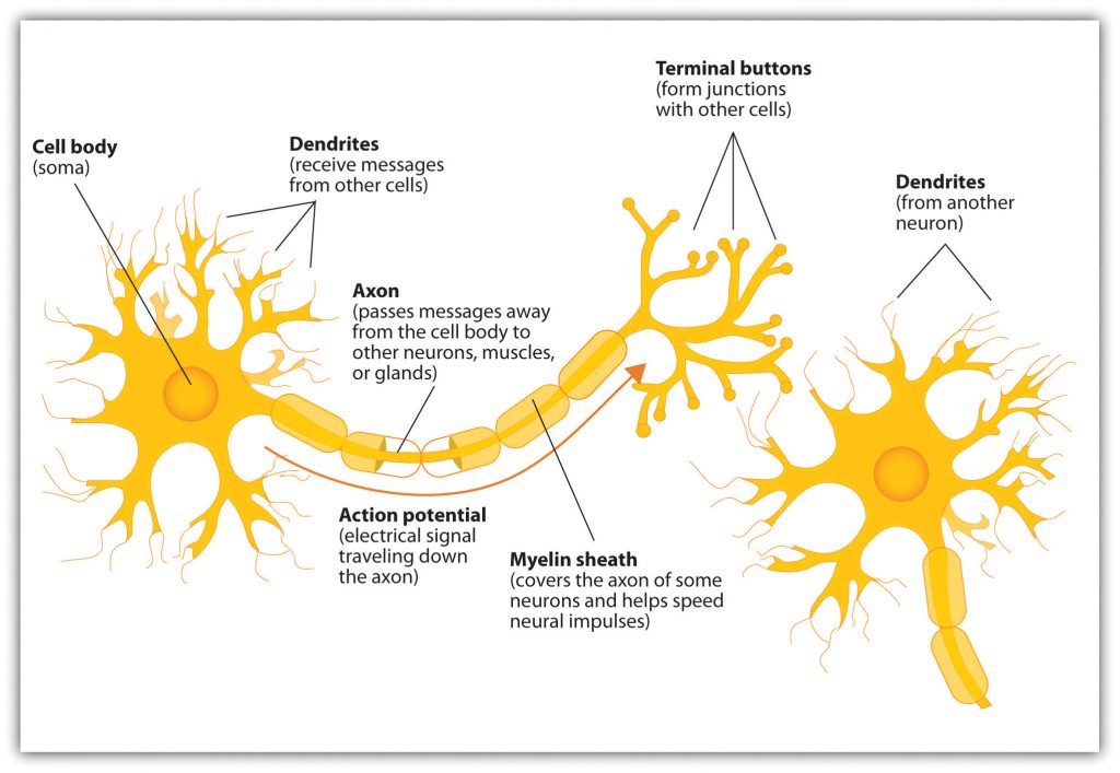
The nervous system is composed of more than 100 billion cells known as neurons. A neuron is a cell in the nervous system whose function is to receive and transmit information. As you can see in the figure, “Components of the Neuron,” neurons are made up of three major parts: a cell body, or soma, which contains the nucleus of the cell and keeps the cell alive; a branching treelike fiber known as the dendrite, which collects information from other cells and sends the information to the soma; and a long, segmented fiber known as the axon, which transmits information away from the cell body toward other neurons or to the muscles and glands.
Components of the Neuron
Neurons, in vitro color
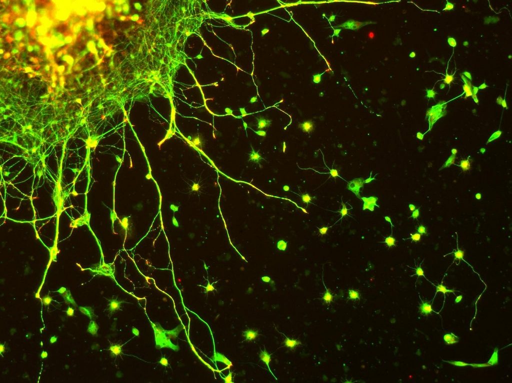
Some neurons have hundreds or even thousands of dendrites, and these dendrites may themselves be branched to allow the cell to receive information from thousands of other cells. The axons are also specialized, and some, such as those that send messages from the spinal cord to the muscles in the hands or feet, may be very long—even up to several feet in length. To improve the speed of their communication, and to keep their electrical charges from shorting out with other neurons, axons are often surrounded by a myelin sheath. The myelin sheath is a layer of fatty tissue surrounding the axon of a neuron that both acts as an insulator and allows faster transmission of the electrical signal. Axons branch out toward their ends, and at the tip of each branch is a terminal button.
Neurons Communicate Using Electricity and Chemicals
The nervous system operates using an electrochemical process. An electrical charge moves through the neuron itself and chemicals are used to transmit information between neurons. Within the neuron, when a signal is received by the dendrites, is it transmitted to the soma in the form of an electrical signal, and, if the signal is strong enough, it may then be passed on to the axon and then to the Terminal buttons (axon terminal containing synaptic vesicles). If the signal reaches the terminal buttons, they are signaled to emit chemicals known as neurotransmitters (chemical messenger of the nervous system), which communicate with other neurons across the spaces between the cells, known as synapses.
This video, Neuron Impulse, shows a model of the electrochemical action of the neuron and neurotransmitters.
The electrical signal moves through the neuron as a result of changes in the electrical charge of the axon. Normally, the axon remains in the resting potential, a state in which the interior of the neuron contains a greater number of negatively charged ions than does the area outside the cell. When the segment of the axon that is closest to the cell body is stimulated by an electrical signal from the dendrites, and if this electrical signal is strong enough that it passes a certain level or threshold, the cell membrane in this first segment opens its gates, allowing positively charged sodium ions that were previously kept out to enter. This change in electrical charge that occurs in a neuron when a nerve impulse is transmitted is known as the action potential. Once the action potential occurs, the number of positive ions exceeds the number of negative ions in this segment, and the segment temporarily becomes positively charged.
Between signals, the neuron membrane’s potential is held in a state of readiness, called the resting potential. Like a rubber band stretched out and waiting to spring into action, ions line up on either side of the cell membrane, ready to rush across the membrane when the neuron goes active and the membrane opens its gates (i.e., a sodium-potassium pump that allows movement of ions across the membrane). Ions in high-concentration areas are ready to move to low-concentration areas, and positive ions are ready to move to areas with a negative charge.
In the resting state, sodium (Na+) is at higher concentrations outside the cell, so it will tend to move into the cell. Potassium (K+), on the other hand, is more concentrated inside the cell and will tend to move out of the cell. In addition, the inside of the cell is slightly negatively charged compared to the outside. This provides an additional force on sodium, causing it to move into the cell.
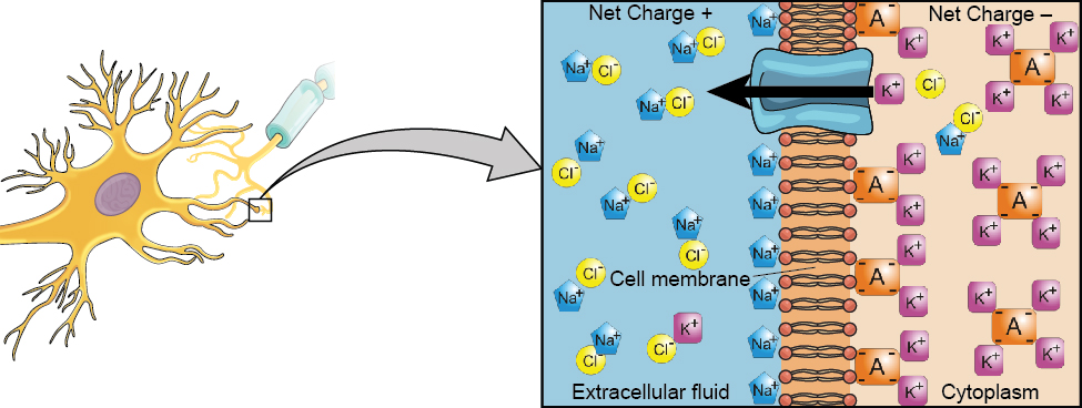
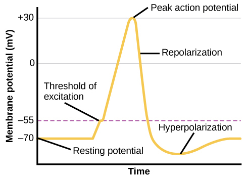
From this resting potential state, the neuron receives a signal and its state changes abruptly. When a neuron receives signals at the dendrites—due to neurotransmitters from an adjacent neuron binding to its receptors (protein on the cell surface where neurotransmitters attach)—small pores, or gates, open on the neuronal membrane, allowing Na+ ions, propelled by both charge and concentration differences, to move into the cell. With this influx of positive ions, the internal charge of the cell becomes more positive. If that charge reaches a certain level, called the threshold of excitation (level of charge in the membrane that causes the neuron to become active), the neuron becomes active and the action potential begins.
Many additional pores open, causing a massive influx of Na+ ions and a huge positive spike in the membrane potential, the peak action potential. At the peak of the spike, the sodium gates close and the potassium gates open. As positively charged potassium ions leave, the cell quickly begins repolarization. At first, it hyperpolarizes, becoming slightly more negative than the resting potential, and then it levels off, returning to the resting potential.
As you can see in the figure “The Myelin Sheath and the Nodes of Ranvier,” the axon is segmented by a series of breaks between the sausage-like segments of the myelin sheath. Each of these gaps is a node of Ranvier. The electrical charge moves down the axon from segment to segment, in a set of small jumps, moving from node to node. When the action potential occurs in the first segment of the axon, it quickly creates a similar change in the next segment, which then stimulates the next segment and so forth as the positive electrical impulse continues all the way down to the end of the axon. As each new segment becomes positive, the membrane in the prior segment closes up again, and the segment returns to its negative resting potential. In this way the action potential is transmitted along the axon, toward the terminal buttons. The entire response along the length of the axon is very fast—it can happen up to 1,000 times each second.
The Myelin Sheath and the Nodes of Ranvier

An important aspect of the action potential is that it operates in an all-or-nothing manner. What this means is that the neuron either fires completely, such that the action potential moves all the way down the axon, or it does not fire at all. Thus neurons can provide more energy to the neurons down the line by firing faster but not by firing more strongly. Furthermore, the neuron is prevented from repeated firing by the presence of a refractory period—a brief time after the firing of the axon in which the axon cannot fire again because the neuron has not yet returned to its resting potential.
Neurotransmitters: The Body’s Chemical Messengers
Not only do the neural signals travel via electrical charges within the neuron, but they also travel via chemical transmission between the neurons. Neurons are separated by junction areas known as synapses, areas where the terminal buttons at the end of the axon of one neuron nearly, but don’t quite, touch the dendrites of another. The synapses provide a remarkable function because they allow each axon to communicate with many dendrites in neighboring cells. Because a neuron may have synaptic connections with thousands of other neurons, the communication links among the neurons in the nervous system allow for a highly sophisticated communication system.
When the electrical impulse from the action potential reaches the end of the axon, it signals the terminal buttons to release neurotransmitters into the synapse. A neurotransmitter is a chemical that relays signals across the synapses between neurons. Neurotransmitters travel across the synaptic space between the terminal button of one neuron and the dendrites of other neurons, where they bind to the dendrites in the neighboring neurons. Furthermore, different terminal buttons release different neurotransmitters, and different dendrites are particularly sensitive to different neurotransmitters. The dendrites will admit the neurotransmitters only if they are the right shape to fit in the receptor sites on the receiving neuron. For this reason, the receptor sites and neurotransmitters are often compared to a lock and key (Figure 3.5 “The Synapse”).
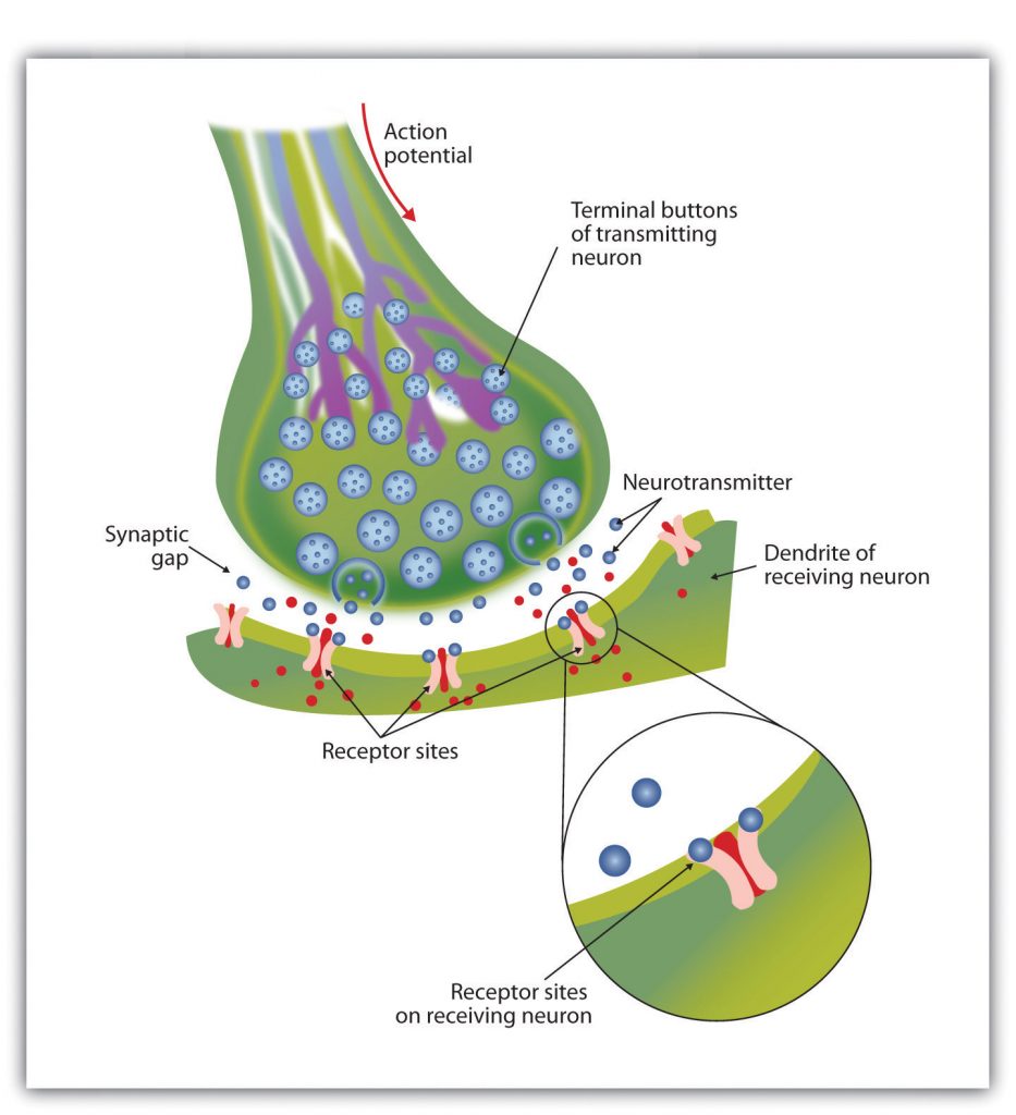
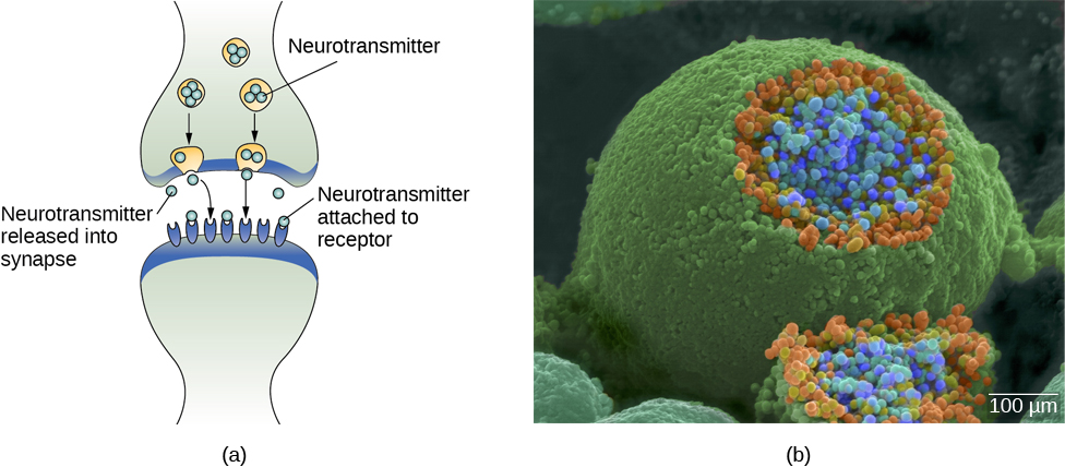
When neurotransmitters are accepted by the receptors on the receiving neurons their effect may be either excitatory (i.e., they make the cell more likely to fire) or inhibitory (i.e., they make the cell less likely to fire). Furthermore, if the receiving neuron is able to accept more than one neurotransmitter, then it will be influenced by the excitatory and inhibitory processes of each. If the excitatory effects of the neurotransmitters are greater than the inhibitory influences of the neurotransmitters, the neuron moves closer to its firing threshold, and if it reaches the threshold, the action potential and the process of transferring information through the neuron begins.
Neurotransmitters that are not accepted by the receptor sites must be removed from the synapse in order for the next potential stimulation of the neuron to happen. This process occurs in part through the breaking down of the neurotransmitters by enzymes, and in part through reuptake, a process in which neurotransmitters that are in the synapse are reabsorbed into the transmitting terminal buttons, ready to again be released after the neuron fires.
More than 100 chemical substances produced in the body have been identified as neurotransmitters, and these substances have a wide and profound effect on emotion, cognition, and behavior. Neurotransmitters regulate our appetite, our memory, our emotions, as well as our muscle action and movement. And as you can see in Table 3.1 “The Major Neurotransmitters and Their Functions,” some neurotransmitters are also associated with psychological and physical diseases.
Drugs that we might ingest—either for medical reasons or recreationally—can act like neurotransmitters to influence our thoughts, feelings, and behavior. An agonist is a drug that has chemical properties similar to a particular neurotransmitter and thus mimics the effects of the neurotransmitter. When an agonist is ingested, it binds to the receptor sites in the dendrites to excite the neuron, acting as if more of the neurotransmitter had been present. As an example, cocaine is an agonist for the neurotransmitter dopamine. Because dopamine produces feelings of pleasure when it is released by neurons, cocaine creates similar feelings when it is ingested. An antagonist is a drug that reduces or stops the normal effects of a neurotransmitter. When an antagonist is ingested, it binds to the receptor sites in the dendrite, thereby blocking the neurotransmitter. As an example, the poison curare is an antagonist for the neurotransmitter acetylcholine. When the poison enters the brain, it binds to the dendrites, stops communication among the neurons, and usually causes death. Still other drugs work by blocking the reuptake of the neurotransmitter itself—when reuptake is reduced by the drug, more neurotransmitter remains in the synapse, increasing its action.
Certain symptoms of schizophrenia are associated with overactive dopamine neurotransmission. The antipsychotics used to treat these symptoms are antagonists for dopamine—they block dopamine’s effects by binding its receptors without activating them. Thus, they prevent dopamine released by one neuron from signaling information to adjacent neurons.
In contrast to agonists and antagonists, which both operate by binding to receptor sites, reuptake inhibitors prevent unused neurotransmitters from being transported back to the neuron. This leaves more neurotransmitters in the synapse for a longer time, increasing its effects. Depression, which has been consistently linked with reduced serotonin levels, is commonly treated with selective serotonin reuptake inhibitors (SSRIs). By preventing reuptake, SSRIs strengthen the effect of serotonin, giving it more time to interact with serotonin receptors on dendrites. Common SSRIs on the market today include Prozac, Paxil, and Zoloft. The drug LSD is structurally very similar to serotonin, and it affects the same neurons and receptors as serotonin. Psychotropic drugs are not instant solutions for people suffering from psychological disorders. Often, an individual must take a drug for several weeks before seeing improvement, and many psychoactive drugs have significant negative side effects. Furthermore, individuals vary dramatically in how they respond to the drugs. To improve chances for success, it is not uncommon for people receiving pharmacotherapy to undergo psychological and/or behavioral therapies as well. Some research suggests that combining drug therapy with other forms of therapy tends to be more effective than any one treatment alone (for one such example, see March et al., 2007).
Test Your Understanding
Key Takeaways
- The central nervous system (CNS) is the collection of neurons that make up the brain and the spinal cord.
- The peripheral nervous system (PNS) is the collection of neurons that link the CNS to our skin, muscles, and glands.
- Neurons are specialized cells, found in the nervous system, which transmit information. Neurons contain a dendrite, a soma, and an axon.
- Some axons are covered with a fatty substance known as the myelin sheath, which surrounds the axon, acting as an insulator and allowing faster transmission of the electrical signal.
- The dendrite is a treelike extension that receives information from other neurons and transmits electrical stimulation to the soma.
- The axon is an elongated fiber that transfers information from the soma to the terminal buttons.
Review Questions
cell in the nervous system whose function is to receive and transmit information
contains the nucleus of the cell and keeps the cell alive
collects information from other cells and sends the information to the soma
transmits information away from the cell body toward other neurons or to the muscles and glands
a layer of fatty tissue surrounding the axon of a neuron that both acts as an insulator and allows faster transmission of the electrical signal.
axon terminal containing synaptic vesicles
chemical messenger of the nervous system
spaces between the cells
a state in which the interior of the neuron contains a greater number of negatively charged ions than does the area outside the cell
change in electrical charge that occurs in a neuron when a nerve impulse is transmitted
protein on the cell surface where neurotransmitters attach
level of charge in the membrane that causes the neuron to become active
a series of breaks between the sausage-like segments of the myelin sheath
areas where the terminal buttons at the end of the axon of one neuron nearly, but don’t quite, touch the dendrites of another.
a chemical that relays signals across the synapses between neurons.
a process in which neurotransmitters that are in the synapse are reabsorbed into the transmitting terminal buttons, ready to again be released after the neuron fires
a drug that has chemical properties similar to a particular neurotransmitter and thus mimics the effects of the neurotransmitter
a drug that reduces or stops the normal effects of a neurotransmitter

