Chapter 8. Tissue Structure and Functions
Ayda Basgul Martin
Unit Outline
Part 1. Tissue: A Higher Level of Organization
- General Structure of Epithelial Tissue
- General Functions of Epithelial Tissue
- Classification of Epithelial Tissue
Learning Objectives
At the end of this unit, you should be able to:
I. Define tissue and describe the importance of tissue level organization to an organism.
II. Describe the structure and function of epithelial, connective, muscle, and nervous tissue.
III. Identify the distinguishing characteristics of these tissues.
IV. Give examples to most common locations of these tissues.
V. Explain the relationships between the structure and function of tissues.
The body contains at least 200 distinct cell types. These cells contain essentially the same internal structures, yet they vary enormously in shape and function. The different types of cells are not randomly distributed throughout the body; rather, they occur in organized layers, a level of organization referred to as tissue.
Part 1: Tissue: A Higher Level of Organization
The term tissue is used to describe a group of cells found together in the body and serves a common function. The cells within a tissue share a common embryonic origin. Microscopic observation reveals that the cells in a tissue share morphological features and are arranged in an orderly pattern that achieves the tissue’s functions. From the evolutionary perspective, tissues appear in more complex organisms. For example, multicellular protists, ancient eukaryotes, do not have cells organized into tissues. Having tissue-level organization increases the efficiency of the body, as different shapes and internal structures are better suited to carry out different functions. Having different tissues for different functions allows for a greater speed of activity and greater effectiveness in performing the various activities.
Although there are many types of cells in the human body, they are organized into four major categories of tissues: epithelial, connective, muscle, and nervous. Each of these categories is characterized by specific functions that contribute to the overall health and maintenance of the body. A disruption of the structure is a sign of injury or disease. Such changes can be detected through histology, the microscopic study of tissue appearance, organization, and function.
The Four Types of Tissues
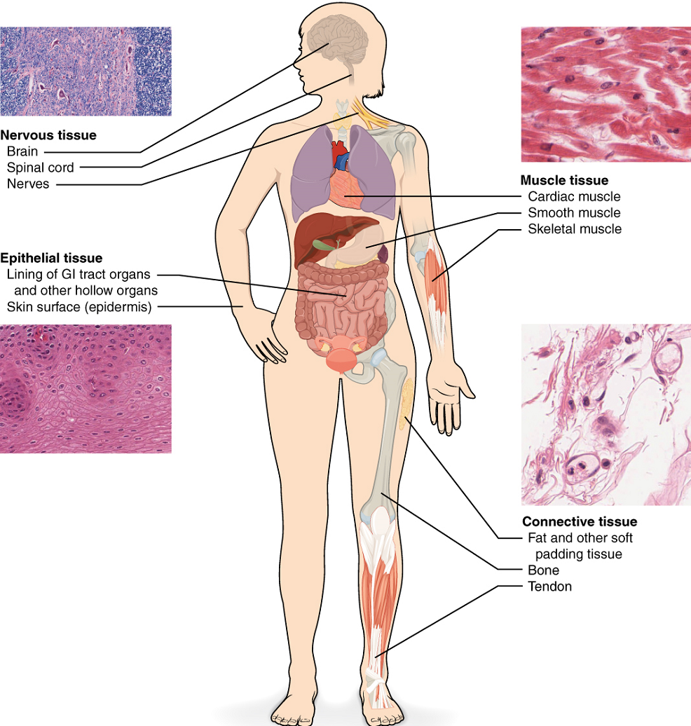
Epithelial tissue, also referred to as epithelium, refers to the sheets of cells that cover exterior surfaces of the body, lines internal cavities and passageways, and forms certain glands. Connective tissue, as its name implies, binds the cells and organs of the body together and functions in the protection, support, and integration of all parts of the body. Muscle tissue is excitable, responding to stimulation and contracting to provide movement, and occurs as three major types: skeletal (voluntary) muscle, smooth muscle, and cardiac muscle in the heart. Nervous tissue is also excitable, allowing the propagation of electrochemical signals in the form of nerve impulses that communicate between different regions of the body (Figure 1).
The next level of organization is the organ, where several types of tissues come together to form a working unit. Just as knowing the structure and function of cells helps you in your study of tissues, knowledge of tissues will help you understand how organs function. The epithelial and connective tissues are discussed in detail in this chapter. Muscle and nervous tissues will be discussed only briefly in this section.
Test Your Knowledge—Part 1
I. Define tissue and describe the importance of tissue-level organization to an organism.
- What is a tissue?
- What is the main benefit to humans of having tissue-level organization?
II. Describe the structure and function of epithelial, connective, muscle, and nervous tissue.
- Describe the general structure and distinguishing characteristics of each of the following:
- Epithelial tissue
- Connective tissue
- Muscle tissue
- Nervous tissue
- Describe the general function of each of the following:
- Epithelial tissue
- Connective tissue
- Muscle tissue
- Nervous tissue
Part 2: Epithelial Tissue
Most epithelial tissues are essentially large sheets of cells covering all the surfaces of the body exposed to the outside world and lining the outside organs and the body cavities. The epithelium also forms much of the glandular tissue of the body. Skin is not the only area of the body exposed to the outside. Other areas include the airways, the digestive tract, as well as the urinary and reproductive systems, all of which are lined by an epithelium. One of the differences between skin epithelia and the epithelia covering the orifices of the body is whether or not it has a thick keratinized layer over it. Hollow organs and body cavities that do not connect to the exterior of the body, which includes blood vessels and serous membranes, are lined by endothelium (plural = endothelia), which is a type of epithelium.
Distinguishing Characteristics of Epithelial Tissue Orifices
All epithelia share some important structural and functional features. This tissue is highly cellular, with little or no extracellular material present between cells. The epithelial cells exhibit polarity with differences in structure and function between the exposed or apical-facing surface of the cell and the basal surface close to the underlying body structures. Particular structures found in some epithelial cells are an adaptation to specific functions. Certain organelles are segregated to the basal sides, whereas other organelles and extensions, such as cilia, when present, are on the apical surface. The basal lamina, a mixture of glycoproteins and collagen, provides an attachment site for the epithelium, separating it from underlying connective tissue. The basal lamina attaches to a reticular lamina, which is secreted by the underlying connective tissue, forming a basement membrane that helps hold it all together.
Epithelial tissues are nearly completely avascular. For instance, no blood vessels cross the basement membrane to enter the tissue, and nutrients must come by diffusion or absorption from underlying tissues or the surface.
Many epithelial tissues are capable of rapidly replacing damaged and dead cells. Sloughing off of damaged or dead cells is a characteristic of surface epithelium and allows our airways and digestive tracts to rapidly replace damaged cells with new cells.
General Functions of Epithelial Tissue
The general functions of the epithelial tissues can be summarized as protection, absorption, secretion and excretion. Epithelial tissues provide the body’s first line of protection from physical, chemical, and biological wear and tear. The cells of an epithelium act as gatekeepers of the body controlling permeability and allowing selective transfer of materials across a physical barrier. All substances that enter the body must cross an epithelium. Some epithelia often include structural features that allow the selective transport of molecules and ions across their cell membranes.
Many epithelial cells are capable of secretion and releasing mucous and specific chemical compounds onto their apical surfaces. The epithelium of the small intestine release digestive enzymes, for example. Cells lining the respiratory tract secrete mucous that traps incoming microorganisms and particles. A glandular epithelium contains many secretory cells.
Classification of Epithelial Tissues
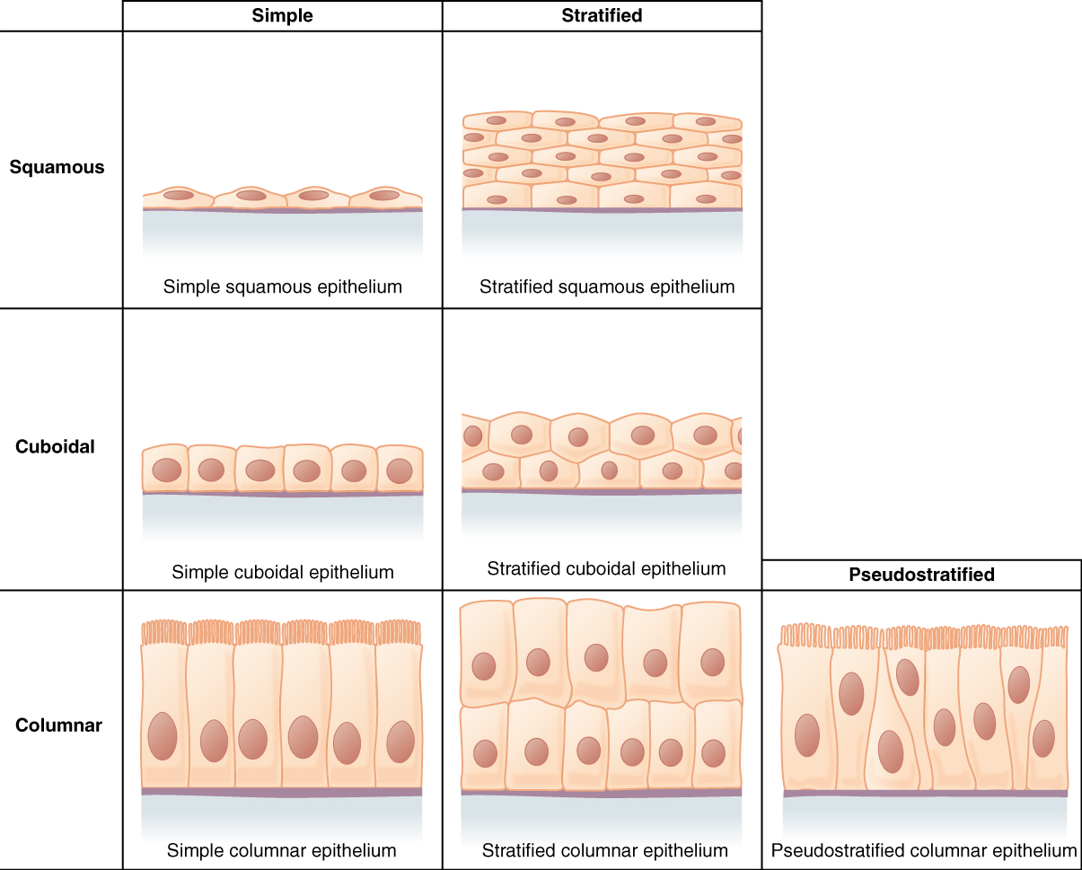
Epithelial tissues are classified according to the shape of the cells and the number of cell layers formed (Figure 2). Cell shapes can be squamous (flattened and thin), cuboidal (boxy, as wide as it is tall), or columnar (rectangular, taller than it is wide). Similarly, the number of cell layers in the tissue can be one—where every cell rests on the basal lamina—which is a simple epithelium, or more than one, which is a stratified epithelium, and only the basal layer of cells rests on the basal lamina. Pseudostratified (pseudo- = “false”) describes tissue with a single layer of irregularly shaped cells that give the appearance of more than one layer. Transitional describes a form of specialized stratified epithelium in which the shape of the cells can vary.
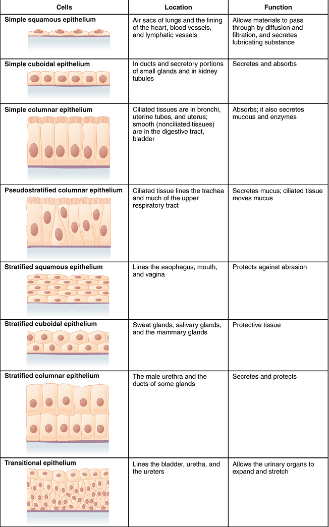
Simple Epithelium: The shape of the cells in the single-cell layer of simple epithelium reflects the functioning of those cells. The cells in simple squamous epithelium have the appearance of thin scales. Squamous cell nuclei tend to be flat, horizontal, and elliptical, mirroring the form of the cell. Simple squamous epithelium, because of the thinness of the cell, is present where the rapid passage of chemical compounds is observed. The alveoli of the lungs where gases diffuse, glomeruli and Bowman’s capsule of the kidney to filter the blood, and the lining of capillaries to allow rapid diffusion of the substances are also made of simple squamous epithelial tissue.
In simple cuboidal epithelium, the nucleus of the box-like cells appears round and is generally located near the center of the cell. These epithelia are active in the secretion and absorption of molecules. Simple cuboidal epithelia are observed in the lining of the kidney tubules and in the ducts of glands (Figure 3).
In simple columnar epithelium, the nucleus of the tall column-like cells tends to be elongated and located in the basal end of the cells (Figure 3). Like the cuboidal epithelia, this epithelium is active in the absorption and secretion of molecules. Simple columnar epithelium forms the lining of some sections of the digestive system and parts of the female reproductive tract. The ciliated columnar epithelium is composed of simple columnar epithelial cells with cilia on their apical surfaces. These epithelial cells are found in the lining of the uterine tubes and parts of the respiratory system, where the beating of the cilia helps remove particulate matter.
Pseudostratified columnar epithelium is a type of epithelium that appears to be stratified but instead consists of a single layer of irregularly shaped and differently sized columnar cells. In pseudostratified epithelium, nuclei of neighboring cells appear at different levels rather than clustered in the basal end (Figure 3). The arrangement gives the appearance of stratification; but in fact, all the cells are in contact with the basal lamina, although some do not reach the apical surface. The pseudostratified columnar epithelium is found in the respiratory tract, where some of these cells have cilia.
Stratified Epithelium: A stratified epithelium consists of several stacked layers of cells. This epithelium protects against physical and chemical wear and tear. The stratified epithelium is named by the shape of the most apical layer of cells, closest to the free space.
Stratified squamous epithelium is the most common type of stratified epithelium in the human body. The apical cells are squamous, whereas the basal layer contains either columnar or cuboidal cells. The top layer may be covered with dead cells filled with keratin. Mammalian skin is an example of this dry, keratinized, stratified squamous epithelium. The lining of the mouth cavity is an example of a nonkeratinized, stratified squamous epithelium. Stratified cuboidal epithelium and stratified columnar epithelium can also be found in certain glands and ducts but are uncommon in the human body (Figure 3).

Test Your Knowledge—Part 2
- Describe the functions of the epithelial tissues.
- Define the criteria to name the type of epithelial tissues.
- Use annotated diagrams to describe the structure and function of each of the eight types of epithelial tissues, and give an example to their common locations.
-
- Simple squamous epithelium.
- Simple cuboidal epithelium.
- Simple columnar epithelium.
- Pseudostratified columnar epithelium.
- Stratified squamous epithelium.
- Stratified cuboidal epithelium.
- Stratified columnar epithelium.
- Transitional epithelium.
4. Compare and contrast the structure and function of:
-
- Simple squamous epithelium and stratified squamous epithelium.
- Simple squamous epithelium and simple columnar epithelium.
- Simple squamous epithelium and simple cuboidal epithelium.
- Simple cuboidal epithelium and simple columnar epithelium.
- Simple cuboidal epithelium and stratified cuboidal epithelium.
Part 3: Connective Tissue
The General Structure of Connective Tissue
As may be obvious from its name, one of the major functions of connective tissue is to connect tissues and organs. Unlike epithelial tissue, which is composed of cells closely packed with little or no extracellular space in between, connective tissue cells are dispersed in a matrix. The matrix usually includes a large amount of extracellular material produced by the connective tissue cells that are embedded within it. The matrix plays a major role in the functioning of this tissue. The major component of the matrix is a ground substance often crisscrossed by protein fibers. This ground substance is usually a fluid, but it can also be mineralized and solid, as in bones. Connective tissues come in a vast variety of forms, yet they typically have in common three characteristic components: cells, large amounts of amorphous ground substance, and protein fibers. The amount and structure of each component correlate with the function of the tissue, from the rigid ground substance in bones supporting the body to the inclusion of specialized cells; for example, a phagocytic cell that engulfs pathogens and also rids tissue of cellular debris.
Functions of Connective Tissues
Connective tissues perform many functions in the body, but most importantly, they support and connect other tissues; from the connective tissue sheath that surrounds muscle cells to the tendons that attach muscles to bones and to the skeleton that supports the positions of the body. Protection is another major function of connective tissue, in the form of fibrous capsules and bones that protect delicate organs and, of course, the skeletal system. Specialized cells in connective tissue defend the body from microorganisms that enter the body. Transport of fluid, nutrients, waste, and chemical messengers is ensured by specialized fluid connective tissues, such as blood and lymph. Adipose cells store surplus energy in the form of fat and contribute to the thermal insulation of the body.
Classification of Connective Tissue
The three broad categories of connective tissue are classified according to the characteristics of their ground substance and the types of fibers found within the matrix (Table 1). Connective tissue proper includes loose connective tissue and dense connective tissue. Both tissues have a variety of cell types and protein fibers suspended in a viscous ground substance. Dense connective tissue is reinforced by bundles of fibers that provide tensile strength, elasticity, and protection. In loose connective tissue, the fibers are loosely organized, leaving large spaces in between. Supportive connective tissue—bone and cartilage—provide structure and strength to the body and protect soft tissues. A few distinct cell types and densely packed fibers in a matrix characterize these tissues. In bone, the matrix is rigid and described as calcified because of the deposited calcium salts. In fluid connective tissue—lymph and blood—various specialized cells circulate in a watery fluid containing salts, nutrients, and dissolved proteins.
| Connective tissue proper | Supportive connective tissue | Fluid connective tissue |
|---|---|---|
Loose connective tissue
|
Cartilage
|
Blood |
Dense connective tissue
|
Bones
|
Lymph |
Connective Tissue Proper
Fibroblasts are present in all connective tissue proper (Figure 4). Fibroblasts are the most abundant cells in connective tissue proper. Fibrocytes, adipocytes, and mesenchymal cells are fixed cells, which means they remain within the connective tissue. Other cells move in and out of the connective tissue in response to chemical signals. Macrophages, mast cells, lymphocytes, plasma cells, and phagocytic cells are found in connective tissue proper but are actually part of the immune system protecting the body.
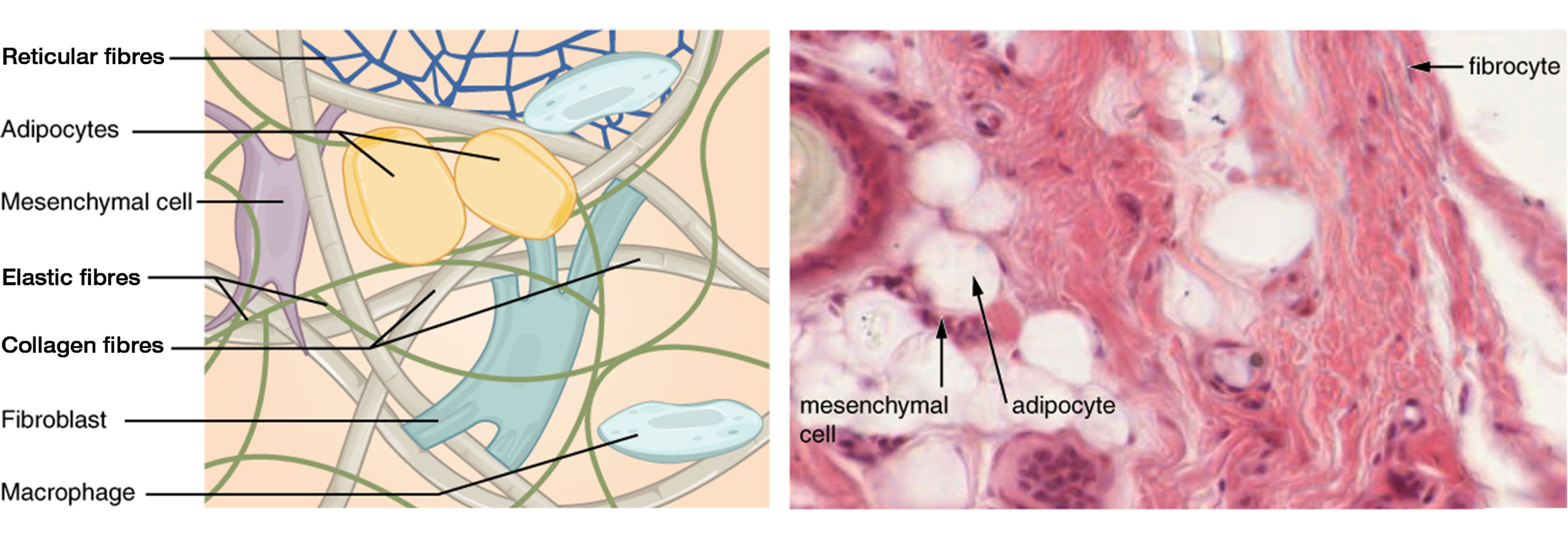
Adipocytes are cells that store lipids as droplets that fill most of the cytoplasm. The mesenchymal cell is a multipotent adult stem cell. These cells can differentiate into any type of connective tissue cells needed for the repair and healing of damaged tissue. The macrophage is a large type of blood cell, which enters the connective tissue matrix from the blood vessels. The macrophage cells are an essential component of the immune system, which is the body’s defense against potential pathogens and degraded host cells. The mast cell found in connective tissue proper, when irritated or damaged, releases histamine, which causes vasodilation and increased blood flow at a site of injury or infection, along with itching, swelling, and redness you recognize as an allergic response.
Three main types of fibers are secreted by fibroblasts: collagen fibers, elastic fibers, and reticular fibers. Collagen fibers, while flexible, have great tensile strength, resist stretching, and give ligaments and tendons their characteristic resilience and strength. These fibers hold connective tissues together, even during the movement of the body. Elastic fibers after being stretched or compressed will return to its original shape. Elastic fibers are prominent in elastic tissues found in skin and the elastic ligaments of the vertebral column. Reticular fibers are narrow and are arrayed in a branching network. They are found throughout the body but are most abundant in the reticular tissue of soft organs, such as liver and spleen, where they anchor and provide structural support to the parenchyma (the functional cells, blood vessels, and nerves of the organ). All of these fiber types are embedded in ground substance, a clear, viscous, colorless matrix made of polysaccharides and proteins, forming the extracellular matrix.
Loose Connective Tissue
Loose connective tissue is found between many organs where it acts both to absorb shock and bind tissues together. It allows water, salts, and various nutrients to diffuse through adjacent or embedded cells and tissues.
Areolar tissue shows little specialization. It contains all the cell types and fibers previously described and is distributed in a random, web-like fashion. It fills the spaces between muscle fibers, surrounds blood and lymph vessels, and supports organs in the abdominal cavity. Areolar tissue underlies most epithelia and represents the connective tissue component of epithelial membranes, which are described further in a later section.
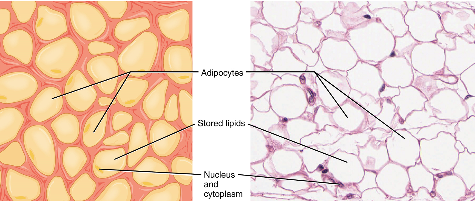
Adipose tissue consists mostly of fat storage cells, with little extracellular matrix (Figure 5). A large number of capillaries allow rapid storage and mobilization of lipid molecules. Fat contributes mostly to lipid storage and can serve as insulation from cold temperatures and mechanical injuries.
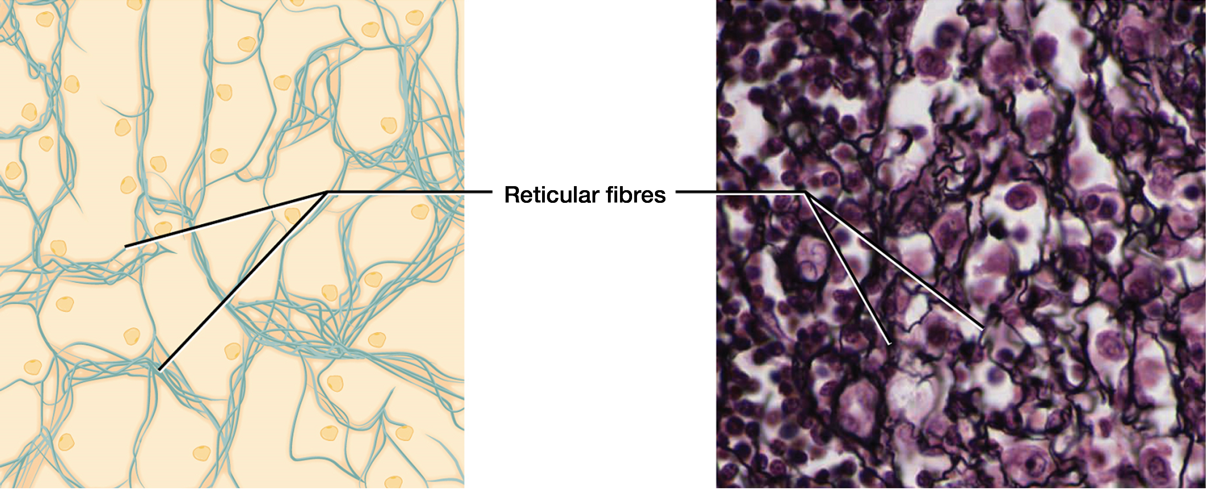
Reticular tissue is a mesh-like, supportive framework for soft organs such as lymphatic tissue, the spleen, and the liver (Figure 6). Reticular cells produce the reticular fibers that form the network to which other cells attach. It derives its name from the Latin reticulus, which means “little net.”
Dense Connective Tissue
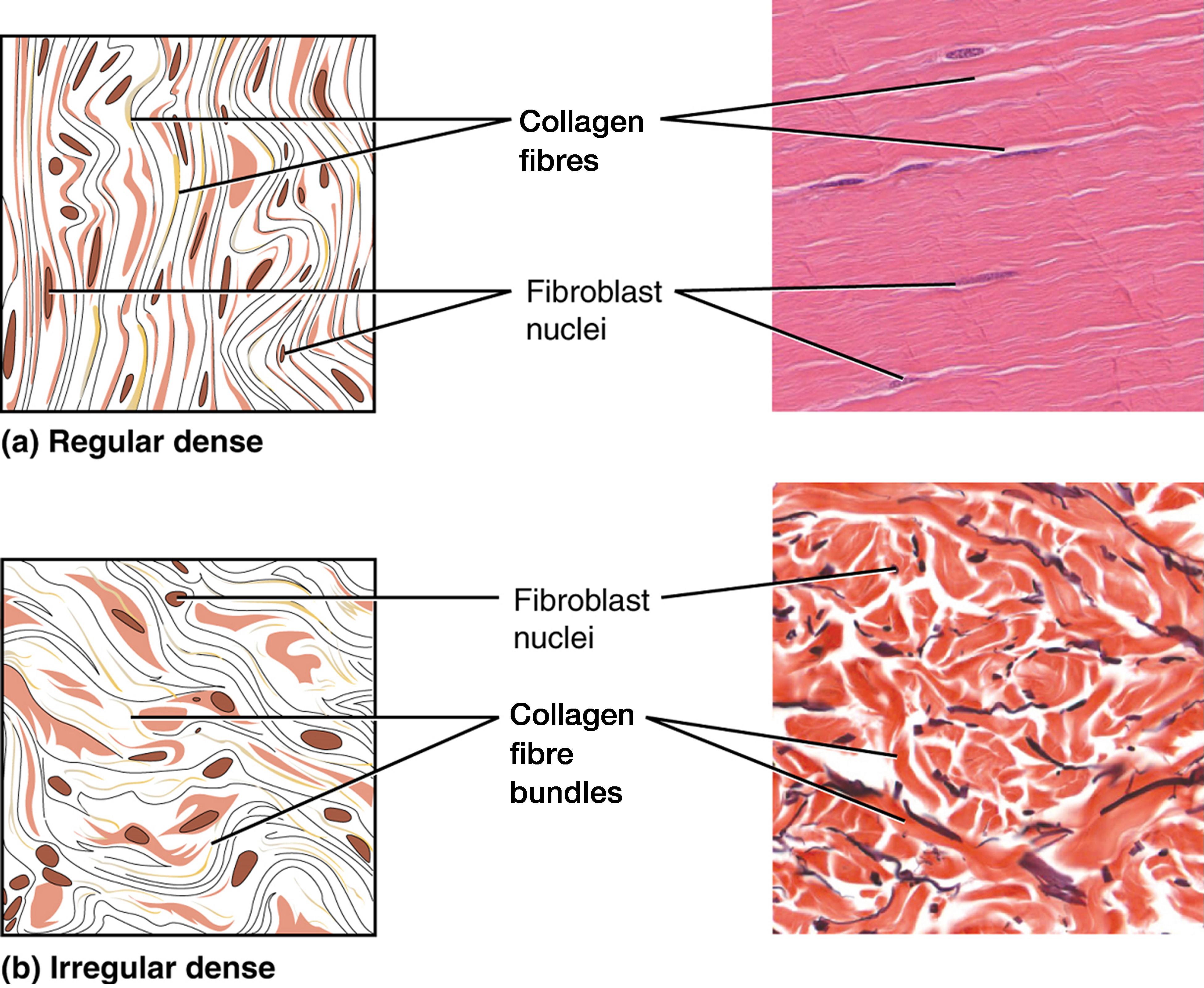
Dense connective tissue (also called fibrous connective tissue) contains more collagen fibers than loose connective tissue. As a consequence, it displays greater resistance to stretching. There are two major categories of dense connective tissue: regular and irregular. Dense regular connective tissue fibers are parallel to each other, enhancing tensile strength and resistance to stretching in the direction of the fiber orientations. Ligaments and tendons are made of dense regular connective tissue, but in ligaments, not all fibers are parallel. Dense regular elastic connective tissue contains elastin fibers in addition to collagen fibers, which allows the ligament to return to its original length after stretching. The ligaments in the vocal folds and between the vertebrae in the vertebral column are elastic.
In dense irregular connective tissue, the direction of fibers is random. This arrangement gives the tissue greater strength in all directions and less strength in one particular direction. In some tissues, fibers crisscross and form a mesh. In other tissues, stretching in several directions is achieved by alternating layers where fibers run in the same orientation in each layer, and it is the layers themselves that are stacked at an angle. The dermis of the skin is an example of dense irregular connective tissue rich in collagen fibers. Dense irregular elastic connective tissue gives arterial walls the strength and the ability to regain their original shape after stretching (Figure 7).
Supportive Connective Tissues
Two major forms of supportive connective tissue, cartilage and bone, allow the body to maintain its posture and protect internal organs.
Cartilage
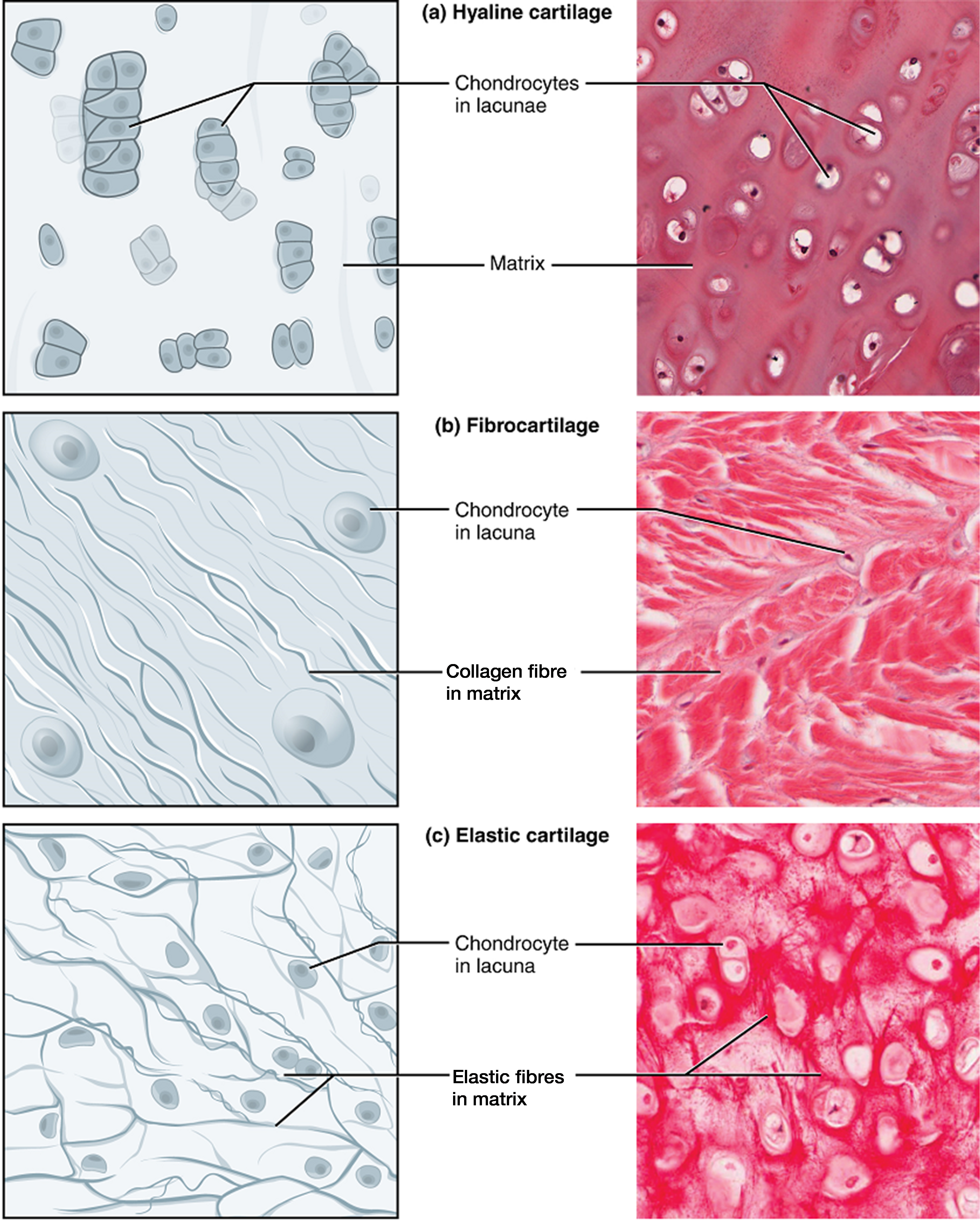
The distinctive appearance of cartilage is due to polysaccharides, which bind with ground substance proteins to form the extracellular matrix. Embedded within the cartilage matrix are chondrocytes, or cartilage cells, and the space they occupy are called lacunae (singular = lacuna). A layer of dense irregular connective tissue, the perichondrium, encapsulates the cartilage. Cartilaginous tissue is avascular, thus all nutrients need to diffuse through the matrix to reach the chondrocytes. This is a factor contributing to the very slow healing of cartilaginous tissues.
The three main types of cartilage tissue are hyaline cartilage, fibrocartilage, and elastic cartilage (Figure 8). Hyaline cartilage, the most common type of cartilage in the body, contains short and dispersed collagen fibers in the matrix. Both strong and flexible, the hyaline cartilage is found in the rib cage and nose and covers bones where they meet to form moveable joints. It makes up a template of the embryonic skeleton before bone formation. A plate of hyaline cartilage at the ends of bone allows continued growth until adulthood. Fibrocartilage is tough because it has thick bundles of collagen fibers dispersed through its matrix. The knee and jaw joints and the intervertebral discs are examples of fibrocartilage. Elastic cartilage contains elastic fibers as well as collagen. This tissue gives rigid support as well as elasticity. Tug gently at your ear lobes, and notice that the lobes return to their initial shape. The external ear contains elastic cartilage.
Bone
Bone is the hardest connective tissue. It provides protection to internal organs and supports the body. Bone’s rigid extracellular matrix contains mostly collagen fibers embedded in a mineralized ground substance containing hydroxyapatite, a form of calcium phosphate. Both components of the matrix, organic and inorganic, contribute to the unusual properties of bone. Without collagen, bones would be brittle and shatter easily. Without mineral crystals, bones would flex and provide little support. Osteocytes, bone cells similar to chondrocytes, are located within lacunae. The histology of transverse tissue from long bone shows a typical arrangement of osteocytes in concentric circles around a central canal. Bone is a highly vascularized tissue. Unlike cartilage, bone tissue can recover from injuries in a relatively short time.
Cancellous bone (“trabecular bone” or “spongy bone”) looks like a sponge under the microscope and contains empty spaces between trabeculae, or arches of bone proper. It is lighter than compact bone and found in the interior of some bones and at the end of long bones. Compact bone is solid and has greater structural strength.
Fluid Connective Tissue
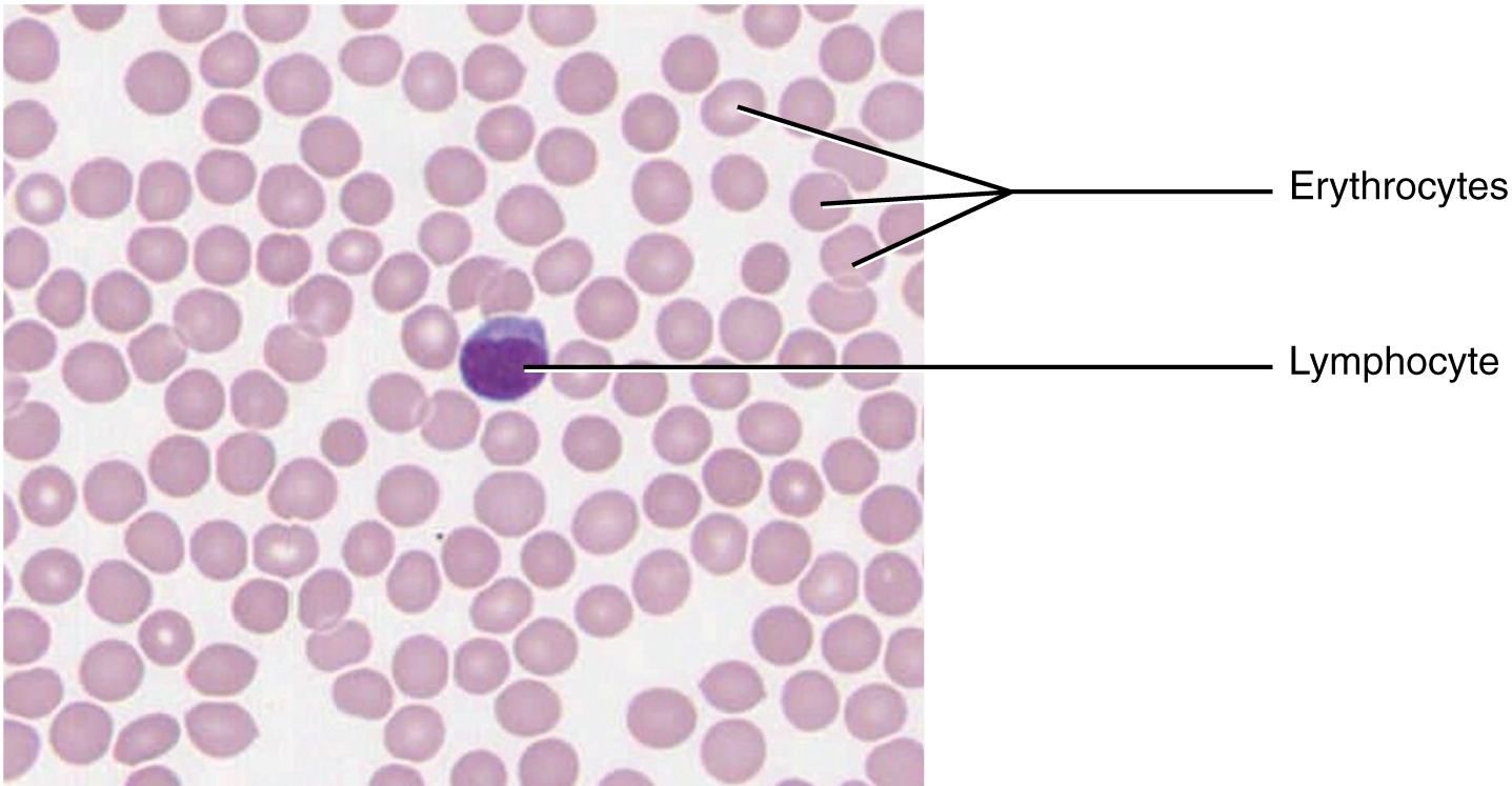
Blood and lymph are fluid connective tissues. Cells circulate in a liquid extracellular matrix. The formed elements circulating in the blood are all derived from hematopoietic stem cells located in bone marrow (Figure 9). Erythrocytes, red blood cells, transport oxygen and some carbon dioxide. Leukocytes, white blood cells, are responsible for defending against potentially harmful microorganisms or molecules. Platelets are cell fragments involved in blood clotting.
Some white blood cells have the ability to cross the endothelial layer that lines blood vessels and enter adjacent tissues. Nutrients, salts, and wastes are dissolved in the liquid matrix and transported through the body.
Lymph contains a liquid matrix and white blood cells. Lymphatic capillaries are extremely permeable, allowing larger molecules and excess fluid from interstitial spaces to enter the lymphatic vessels. Lymph drains into blood vessels, delivering molecules to the blood that could not otherwise directly enter the bloodstream. In this way, specialized lymphatic capillaries transport absorbed fats away from the intestine and deliver these molecules to the blood.
Test Your Knowledge—Part 3
Create a table stating:
-
- The matrix composition,
- The cellular types,
- The main function(s), and
- Specific examples of each of the following types of connective tissue:
-
-
- Fluid connective tissue
- Loose connective tissue
- Dense connective tissue
- Cartilage
- Bone
-
Part 4: Muscle Tissue
Muscle tissue is characterized by properties that allow movement. Muscle cells are excitable; they respond to a stimulus. They are contractile, meaning they can shorten and generate a pulling force. When attached between two movable objects, in other words, bones, contractions of the muscles cause the bones to move. Some muscle movement is voluntary, which means it is under conscious control. For example, a person decides to open a book and read a chapter on anatomy. Other movements are involuntary, meaning they are not under conscious control, such as the contraction of your pupil in bright light. Muscle tissue is classified into three types according to structure and function: skeletal, cardiac, and smooth (Table 2).
| Tissue | Histology | Function | Location |
|---|---|---|---|
| Skeletal | Long cylindrical fiber; striated; many peripherally-located nuclei | Voluntary movement; thermogenesis; organ protection | Attached to bones; found around entrance points to the body (e.g., mouth, anus) |
| Cardiac | Short, branched fibers; striated; single central nucleus | Contracts to pump blood | Heart walls |
| Smooth | Short, spindle-shaped fibers; no evident striation; single nucleus | Involuntary movement; moves material through the digestive tract and ducts; regulates blood flow in arteries | Walls of major organs and passageways |
Skeletal Muscle
Skeletal muscle is attached to bones, and its contraction makes possible locomotion, facial expressions, posture, and other voluntary movements of the body. Forty percent of your body mass is made up of skeletal muscle. Skeletal muscles generate heat as a byproduct of their contraction and thus participate in thermal homeostasis. Shivering is an involuntary contraction of skeletal muscles in response to perceived lower-than-normal body temperature.
The muscle cells, muscle fibers or myocytes, and their numbers remain relatively constant throughout life. Skeletal muscle tissue is arranged in bundles surrounded by connective tissue. Under the light microscope, muscle cells appear striated with many nuclei squeezed along the membranes (Figure 10a). The striation is due to the regular alternation of the contractile proteins actin and myosin, along with the structural proteins that couple the contractile proteins to connective tissues. The cells are multinucleated as a result of the fusion of the many myoblasts that fuse to form each long muscle fiber.
Cardiac Muscle
Cardiac muscle forms the contractile walls of the heart. The cells of cardiac muscle, known as cardiomyocytes, also appear striated under the microscope. Unlike skeletal muscle fibers, cardiomyocytes are single cells typically with a single centrally located nucleus.
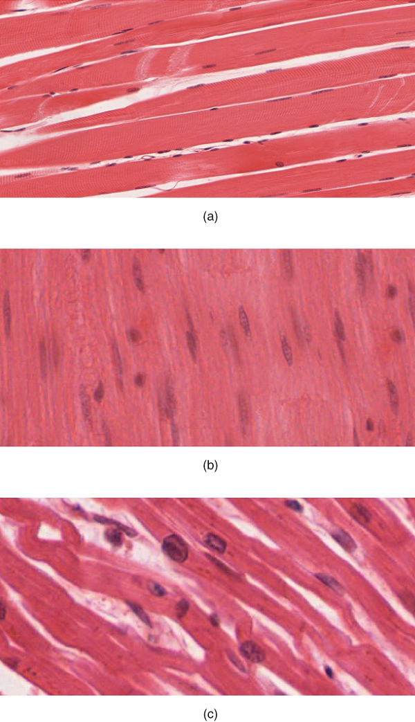
A principal characteristic of cardiomyocytes is that they contract on their own intrinsic rhythms without any external stimulation. Cardiomyocyte attach to one another with specialized cell junctions called intercalated discs. Attached cells form long, branching cardiac muscle fibers that are (Figure 10c), essentially, a mechanical and electrochemical syncytium allowing the cells to synchronize their actions. The cardiac muscle pumps blood through the body and is under involuntary control.
Smooth muscle
Smooth muscle tissue contraction is responsible for involuntary movements in the internal organs. It forms the contractile component of the digestive, urinary, and reproductive systems as well as the airways and arteries. Each cell is spindle shaped with a single nucleus and no visible striations (Figure 10b).

Test Your Knowledge—Part 4
- Compare and contrast the three types of muscle tissue by discussing each of the following characteristics:
- The structure of each of the three types of muscle tissue
- How each type of muscle tissue is controlled (i.e., whether voluntary control is available or not)
- The function(s) of each of the three types of muscle tissue
- Locations of these three types of muscle tissues
Part 5: Nervous Tissue
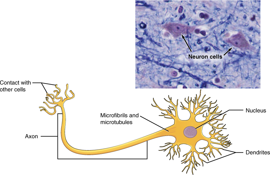
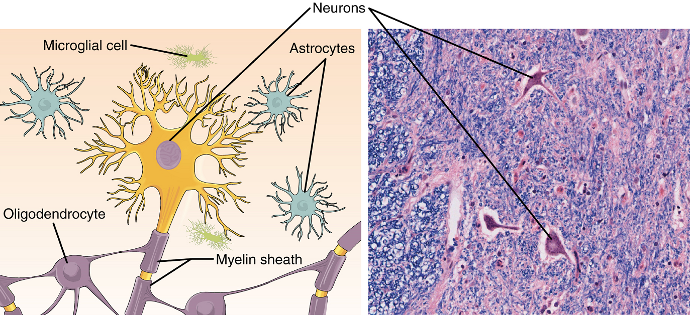
Nervous tissue is characterized as being excitable and capable of sending and receiving electrochemical signals that provide the body with information. Two main classes of cells make up nervous tissue: the neuron and neuroglia (Figure 11). Neurons propagate information via electrochemical impulses, called action potentials, which are biochemically linked to the release of chemical signals. Neuroglia play an essential role in supporting neurons and modulating their information propagation.
Neurons display distinctive morphology, well suited to their role as conducting cells, with three main parts. The cell body includes most of the cytoplasm, the organelles, and the nucleus. Dendrites branch off the cell body and appear as thin extensions. A long “tail,” the axon, extends from the neuron body and can be wrapped in an insulating layer known as myelin, which is formed by accessory cells. The synapse is the gap between nerve cells, or between a nerve cell and its target, for example, a muscle or a gland, across which the impulse is transmitted by chemical compounds known as neurotransmitters.
The second class of neural cells comprises the neuroglia or glial cells (Figure 12), which have been characterized as having a simple support role. The word “glia” comes from the Greek word for glue. Recent research is shedding light on the more complex role of neuroglia in the function of the brain and nervous system.
The presence of the nervous tissue throughout the body and its organization allows it to receive, integrate, and provide information to the entire body. This ensures that appropriate responses can occur among all body systems within an intact organism, both under normal conditions as well as during times of stress.

Test Your Knowledge—Part 5
- Name and describe both of the two main cell types according to their locations in the central and peripheric nervous system. In your description, include:
- Their general cellular morphology (i.e., their shape)
- Their main function(s)
- Their locations
Practice
For the exercise below, drag the answers to the correct empty boxes.
Image Descriptions
Figure 8.1. Four Types of Tissues. The four types of tissues—epithelial, connective, muscle, and nervous tissue—are illustrated in this picture of the human body sketch, and a micrograph was added as a visual example for all four. The brain, spinal cord, and nerves are labeled on the sketch to show the locations of the nervous tissue. Next in the clockwise direction, three types of muscle tissue are listed as the smooth, cardiac, and skeletal muscle tissues and labeled within the sketch as cardiac muscle found in the wall of the heart, smooth muscle on the wall of the stomach, and skeletal muscle found in the left anterior forearm muscles. The connective tissue examples labeled in the illustration are the fat and other soft padding tissue, bone, and tendon. Lastly, the lining of the GI tract organs and other hollow organs and skin surface (epidermis) are labeled in the illustration, and a micrograph is added under epithelial tissue. Clockwise from nervous tissue, LM × 872, LM × 282, LM × 460, LM × 800. (Micrographs provided by the Regents of University of Michigan Medical School © 2012.) [Return to image.]
Figure 8.2. Types of Epithelial Tissue. Epithelial tissues are classified according to how many layers they formed and the shape of the apical layer of cells in the tissue. The function of the epithelial tissue can be classified as protection of underlining tissues, absorption, secretion, and excretion. Epithelial tissues also form the different types of glands. Simple epithelial tissue is organized as a single layer of cells, and stratified epithelial tissue is formed by several layers of cells. Pseudostratified epithelial tissue is a single layer of cells that appear to be multiple layers because of the position of their nuclei. Epithelial tissue is further defined by the shape of the apical layer of cells in the tissue as squamous (flatten and thin), cuboidal (boxy, as wide as it is tall), and columnar (rectangular, taller than it is wide). In this illustration, simple squamous (one layer flat), simple cuboidal, simple columnar, stratified squamous, stratified cuboidal, stratified columnar, and pseudostratified epithelium are presented. [Return to image.]
Figure 8.3. Summary of Epithelial Tissue Types. Different types of epithelial tissue serve different functions and are found in different locations in the body. In this illustrated table, the examples for the locations of the simple squamous epithelium are the air sacs of the lungs, the lining of the heart, blood vessels, and lymphatic vessels, and the functions are allowing materials to pass through by diffusion and filtration and secreting lubricating substances. The example of locations of the simple cuboidal epithelium is the ducts and secretory portions of small glands and kidney tubules, and the listed functions are secretion and absorption. Example of locations of the simple columnar epithelium is ciliated ones in the bronchi, uterine tubes, and uterus; smooth (non-ciliated tissues) are in the digestive tract, bladder, and absorption and secretion of the mucous and enzymes. The pseudostratified columnar epithelium locations are the trachea and much of the upper respiratory tract and secrete mucus and cilia move the mucus. The stratified squamous epithelium lines the esophagus, mouth, and vagina and protects against abrasion. The stratified cuboidal epithelium is found within sweat glands, salivary glands, and mammary glands and serves as protective tissue. The stratified columnar epithelium lines the male urethra and the ducts of some glands and serve to secrete and protect. Lastly, the transitional epithelium lines the bladder, urethra, and ureters and allows the urinary organs to expand and stretch. [Return to image.]
Figure 8.4. Connective Tissue Proper. Connective tissue proper includes both loose (or areolar) connective tissue and dense connective tissue. Loose connective tissue previously included areolar, reticular, and adipose tissues, although this system has been revised to only include areolar tissue. Loose connective tissue generally works to hold organs, anatomic structures, and tissues in place. The extracellular matrix is the most significant feature of loose connective tissue with large spaces between fibers. Dense connective tissue proper is composed of a higher density of fibers, which may be regular (with parallel fibers such as that of tendons and ligaments) or irregular (with multidirectional fibers such as that of the pericardium), or elastic (with significant embedded elastin such as that of arteries). Fibroblasts produce this fibrous tissue. Connective tissue proper includes the fixed cells fibrocytes, adipocytes, and mesenchymal cells. The figure contains an illustration of the connective tissue proper on the left hand with reticular, elastic, and collagen fibers and mesenchymal cells, macrophage, adipocytes, and fibroblast labeled on it. On the right hand, it shows a histologic photo of the tissue with the fibrocyte, adipocytes, and mesenchymal cell labeled. LM × 400. (Micrograph provided by the Regents of the University of Michigan Medical School © 2012.) [Return to image.]
Figure 8.5. Adipose Tissue. Adipose tissue, also known as fat tissue or fatty tissue, is a connective tissue that is mainly composed of fat cells called adipocytes. Adipocytes are energy-storing cells that contain large globules of fat known as lipid droplets surrounded by a structural network of fibers. This is a connective tissue that consists of fat cells with a little extracellular matrix. It stores fat for energy and provides insulation. Adipocytes are categorized into three different cell types—white, brown, and beige adipocytes—based on their origin, location, and function. White adipocytes are the most abundant adipocytes in the human body. They are filled with a large, single lipid droplet and contain few cellular organelles. Brown adipocytes are very metabolically active cells that contain multiple lipid droplets and a high concentration of mitochondria (a cellular organelle that allows brown adipocytes to generate heat). In this figure, there is an illustration on the left and a histologic photo of the adipose tissue on the right with adipocytes, stored lipids, and nucleus and cytoplasm labeled in both. LM × 800. (Micrograph provided by the Regents of the University of Michigan Medical School © 2012.) [Return to image.]
Figure 8.6. Reticular Tissue. This is a loose connective tissue made up of a network of reticular fibers that provides a supportive framework for soft organs. This figure contains an illustration on the left and a photo of the microscopy slide on the right of reticular tissue with reticular fibers labeled in both. Reticular tissue forms the stroma for the spleen, lymph nodes, red bone marrow, liver, and kidneys. Reticular tissue can be considered more of a structural framework for tissues (stroma) than an actual tissue type. Because of this, reticular tissue appears very different from one organ to the next, and reticular fibers are difficult to visualize using normal light microscopy techniques in organs other than lymph nodes. Widely spaced fibroblasts within reticular tissue secrete proteins that assemble into reticular fibers, but they are difficult to identify, since fibroblast nuclei stain the same dark color as reticular fibers when viewed under a microscope. LM × 1600. (Micrograph provided by the Regents of the University of Michigan Medical school 2012.) [Return to image.]
Figure 8.7. Dense Connective Tissue. Dense connective tissue is often seen as the capsules enclosing organs and, in particular, tubular structures, but is most strikingly characterized in its appearance as tendons and ligaments. These are basically dense masses of collagenic fibers and fibroblasts arranged in an orderly manner, with the cells and fibers being oriented in the same direction (i.e., parallel to the long axis of the tendon). Primarily, there is a predominance of fibroblasts, but these secrete increasing amounts of collagen, and the bulk of the tendon becomes fibrous. (a) Dense regular connective tissue consists of collagenous fibers packed into parallel bundles. An illustration on the left and histology slide on the right are shown, and collagen fibers and fibroblast nuclei are labeled (b) Dense irregular connective tissue consists of collagenous fibers interwoven into a mesh-like network shown with an illustration on the left and a photo of microscopy slide on the right with fibroblast nuclei and collagen fiber bundles labeled on. From the top, LM × 1000, LM × 200. (Micrographs provided by the Regents of the University of Michigan Medical School © 2012.) [Return to image.]
Figure 8.8. Types of Cartilage. Cartilage is a connective tissue consisting of collagenous fibers embedded in a firm matrix of chondroitin sulfates. Cartilage is a flexible connective tissue that differs from bone in several ways; it is avascular, and its microarchitecture is less organized than bone. The cells of the cartilage are called chondrocytes, and they are scattered and lie firmly fixed in a matrix supported by collagen and elastic fibers. Cartilage is not innervated and therefore relies on diffusion to obtain nutrients. This causes it to heal very slowly. The main cell types in cartilage are chondrocytes, the ground substance is chondroitin sulfate, and the fibrous sheath is called perichondrium. (a) Hyaline cartilage provides support with some flexibility. The example is from dog tissue. Chondrocytes in lacunae and matrix are labeled. (b) Fibrocartilage provides some compressibility and can absorb pressure. Chondrocyte in lacunae and collagen fiber in matrix are labeled. (c) Elastic cartilage provides firm but elastic support. Chondrocyte in lacunae and elastic fibers in matrix labeled. From the top, LM × 300, LM × 1200, LM × 1016. (Micrographs provided by the Regents of the University of Michigan Medical School © 2012.) [Return to image.]
Figure 8.9. Blood: A Fluid Connective Tissue. Blood is a fluid connective tissue containing erythrocytes and various types of leukocytes that circulate in a liquid extracellular matrix. In this figure, a photo of a microscopy slide shows the peripheric smear of the blood with labels for erythrocytes and a lymphocyte. A peripheric smear or blood smear is a sample of blood that’s spread on a glass slide, which is treated with a special stain. In the past, all blood smears were examined under a microscope by laboratory professionals. Now automated digital systems may be used to help examine blood smears. The purpose of examining a blood smear is to check the size, shape, and number of three types of blood cells: Red blood cells, which carry oxygen throughout the body. White blood cells, which fight infection. Platelets, which help your blood to clot. LM × 1600. (Micrograph provided by the Regents of the University of Michigan Medical School © 2012.) [Return to image.]
Figure 8.10. Muscle Tissue. The illustration is showing the histologic view of the three types of muscle tissue. (a) Skeletal muscle cells have prominent striation and more than one nucleus and nuclei on their periphery; for those reasons, they are called either striated muscle fibers or multinucleated cells. (b) Smooth muscle cells have a single nucleus in the center of the muscle fibers and no visible striations. (c) Cardiac muscle cells appear striated but not as frequent as skeletal muscle; for that reason, they are called semi-striated (partially striated) muscle fibers. Cardiac muscle fibers have a single nucleus, and they are branched. Cardiac muscles have a distinctive feature called intercalated discs. Intercalated discs are complex structures that connect adjacent cardiac muscle cells. From the top, LM × 1600, LM × 1600, LM × 1600. (Micrographs provided by the Regents of the University of Michigan Medical School © 2012.) [Return to image.]
Figure 8.11. The Neuron. Neurons (also called neurons or nerve cells) are the fundamental units of the brain and nervous system, the cells responsible for receiving sensory input from the external world, for sending motor commands to our muscles, and for transforming and relaying the electrical signals to our glands. More than that, their interactions define who we are as people. We have roughly 100 billion neurons that interact closely with other cell types, broadly classified as glia (these may actually outnumber neurons, although it’s not really known). The cell body of a neuron, also called the soma, contains the nucleus and mitochondria. The dendrites transfer the nerve impulse to the soma. The axon carries the action potential away to another excitable cell. This figure contains an illustration of a multipolar neuron and its components (axon, dendrites, microfibrils, microtubules, and nucleus) labeled and a photo of nervous system histology with labeled neurons. A multipolar neuron is a type of neuron that possesses a single axon and many dendrites (and dendritic branches), allowing for the integration of a great deal of information from other neurons. LM × 1600. (Micrograph provided by the Regents of University of Michigan Medical School © 2012.) [Return to image.]
Figure 8.12. Nervous Tissue. Nervous tissue is made up of neurons and neuroglia. The cells of nervous tissue are specialized to transmit and receive impulses. Nervous tissue is found in the brain, spinal cord, and nerves. It is responsible for coordinating and controlling many body activities. It stimulates muscle contraction, creates an awareness of the environment, and plays a major role in emotions, memory, and reasoning. To do all these things, cells in nervous tissue need to be able to communicate with each other by way of electrical nerve impulses. The cells in nervous tissue that generate and conduct impulses are called neurons or nerve cells. These cells have three principal parts: the dendrites, the cell body, and one axon. The main part of the cell, the part that carries on the general functions, is the cell body. Dendrites are extensions, or processes, of the cytoplasm that carry impulses to the cell body. An extension or process called an axon carries impulses away from the cell body. Nervous tissue also includes cells that do not transmit impulses but instead support the activities of the neurons. These are the glial cells (neuroglial cells), together termed the neuroglia. Supporting, or glia, cells bind neurons together and insulate the neurons. Some are phagocytic and protect against bacterial invasion, while others provide nutrients by binding blood vessels to the neurons. This figure contains an illustration of the CNS neuron and neuroglia. The neuron and microglial cell, oligodendrocyte, astrocytes, and myelin sheath are labeled. Also, a photo of nervous system microscopy is presented on the right with labeled neurons. LM × 872. (Micrograph provided by the Regents of University of Michigan Medical School © 2012.) [Return to image.]
Group of many similar cells (though sometimes composed of a few related types) that work together to perform a specific function.
Type of tissue that serves primarily as a covering or lining of body parts, protecting the body; it also functions in absorption, transport, and secretion.
Type of tissue that serves to hold in place, connect, and integrate the body’s organs and systems.
Microscopic study of tissue architecture, organization, and function.
(In physiology) under conscious control of the brain.
Opening such as mouth, nares, anus, etc.
That part of a cell or tissue, which, in general, faces an open space.
That part of a tissue close to underlying body structures.
Thin extracellular layer that lies underneath epithelial cells and separates them from other tissues.
Matrix containing collagen and elastin secreted by connective tissue; a component of the basement membrane.
In epithelial tissue, a thin layer of fibrous material that anchors the epithelial tissue to the underlying connective tissue; made up of the basal lamina and reticular lamina.
Lacking blood vessels.
Atom with an overall positive or negative charge. Many function as electrolytes.
Molecule (usually a protein) that catalyzes chemical reactions.
Tissue that consists of a single layer of flat scale-like cells; promotes diffusion and filtration across surface.
Tissue that consists of a single layer of cube-shaped cells; promotes secretion and absorption in ducts and tubules.
Tissue that consists of a single layer of column-like cells; promotes secretion and absorption in tissues and organs.
Small appendage on certain cells formed by microtubules and modified for movement of materials across the cellular surface (singular = cilium).
(Also, fallopian tube or oviduct) duct that facilitates transport of an ovulated oocyte to the uterus.
Tissue that consists of a single layer of irregularly shaped and sized cells that give the appearance of multiple layers; found in ducts of certain glands and the upper respiratory tract.
Tissue that consists of multiple layers of cells with the most apical being flat scale-like cells; protects surfaces from abrasion.
Type of structural protein that gives skin, hair, and nails its hard, water-resistant properties.
Tissue that consists of two or more layers of cube-shaped cells, found in some ducts.
Tissue that consists of two or more layers of column-like cells, contains glands, and is found in some ducts.
(In connective tissue) extracellular material that is produced by the cells embedded in it, containing ground substance and fibers.
Fluid or semi-fluid portion of the matrix.
Cell process (a form of endocytosis) in which a cell engulfs and ingests another large particle or cell.
An infectious agent that causes disease, typically a bacterium, virus, fungus, or microscopic parasite.
Fluid contained within the lymphatic system, consisting of interstitial fluid, leukocytes (white blood cells), proteins (including antibodies), and fats.
Specialized areolar tissue rich in stored fat.
Connective tissue containing a viscous matrix, fibers, and cells.
Thick consistency between solid and liquid.
Most abundant cell type in connective tissue, secretes protein fibers and matrix into the extracellular space.
Mature, less active form of a fibroblast.
Lipid storage cells.
Embryonic tissue from which connective tissue cells derive.
Ameboid (irregular outline with peripheral projections) phagocyte found in several tissues throughout the body.
Cell found in the skin and the lining of body cells that contains cytoplasmic granules with vasoactive mediators such as histamine.
White blood cell characterized by a large nucleus and small rim of cytoplasm.
Differentiated B cell that is actively secreting antibody.
Describes the condition of being able to differentiate into different types of cells within a given cell lineage or small number of lineages, such as a red blood cell or white blood cell.
Cell that is oligo-, multi-, or pleuripotent that has the ability to produce additional stem cells rather than becoming further specialized.
(In immunology) referring to the organism in, or on, which a pathogen lives.
Vasoactive (active on blood vessels) mediator in granules of mast cells and is the primary cause of allergies and anaphylactic shock.
Opening up, or increasing interior (lumen) diameter of a blood vessel.
The most abundant of three protein fibres found in the extracellular matrix of connective tissues.
Fibrous protein within connective tissue that contains a high percentage of the protein elastin that allows the fibers to stretch and return to original size.
Fine fibrous protein, made of collagen subunits, which cross-link to form supporting “nets” within connective tissue.
Functional cells of a gland or organ, in contrast with the supportive or connective tissue of a gland or organ.
cartilage cells
(Plural = lacunae) small spaces in bone or cartilage tissue that cells occupy.
Layer of dense irregular connective tissue surrounding cartilage.
Most common type of cartilage, smooth and made of short collagen fibers embedded in a chondroitin sulfate ground substance.
Tough form of cartilage, made of thick bundles of collagen fibers embedded in chondroitin sulfate ground substance.
Type of cartilage, with elastin as the major protein, characterized by rigid support as well as elasticity.
A form of calcium phosphate mineral found in bones (also hydroxylapatite).
Primary cell in mature bone; responsible for maintaining the matrix.
Relating to circulation of blood.
(Also, cancellous bone) trabeculated osseous tissue that supports shifts in weight distribution.
(Also, hematopoiesis) production of the formed elements of blood.
Red blood cell.
White blood cell.
(Also, thrombocytes) one of the formed elements of blood that consists of cell fragments broken off from megakaryocytes.
Layer of smooth, simple squamous epithelium that lines the endocardium and blood vessels.
(In physiology) though under nervous control (usually from the brain), control is not conscious.
Usually attached to bone, under voluntary control, each cell is a fiber that is multinucleated and striated.
Heart muscle, under involuntary control, composed of striated cells that attach to form fibres, each cell contains a single nucleus, contracts autonomously.
Under involuntary control, moves internal organs, cells contain a single nucleus, are spindle-shaped, and do not appear striated; each cell is a fiber.
Steady state of body systems that living organisms maintain.
Muscle cell (also muscle fiber).
Alignment of parallel actin and myosin filaments, which form a banded pattern.
A multinucleate cell formed by the fusion of multiple cells or the division of nuclei.
Excitable neural cell that transfer nerve impulses.
Supportive neural cells.
Change in voltage of a cell membrane in response to a stimulus that results in transmission of an electrical signal; unique to neurons and muscle fibres.
Internal material between the cell membrane and nucleus of a cell, mainly consisting of a water-based fluid called cytosol, within which are all the other organelles and cellular solute and suspended materials.
Any of several different types of membrane-enclosed specialized structures in the cell that perform specific functions for the cell.
Cell’s central organelle; contains the cell’s DNA.
One of many branchlike processes that extends from the neuron cell body and functions as a contact for incoming signals (synapses) from other neurons or sensory cells.
Single process of the neuron that carries an electrical signal (action potential) away from the cell body toward a target cell.
Lipid-rich insulating substance surrounding the axons of many neurons, allowing for faster transmission of electrical signals.
Narrow junction across which a chemical signal passes from neuron to the next, initiating a new electrical signal in the target cell.
Chemical signal that is released from the synaptic end bulb of a neuron to cause a change in the target cell.
