Cellular Respiration
Learning Objectives
After completing the lab, the student will be able to:
- Determine the site of respiration in the cell.
Activity 1: Pre-Assessment:
- What function do mitochondria fulfill in the cell? What kind of staining would allow visualization of mitochondrial activity?
- Which plant tissue would you choose to stain mitochondria? Explain your choice.
- Discuss the answers to questions 1 and 2 with the class.
Activity 1: Staining Mitochondria with Janus Green B
You will investigate the site of oxidative phosphorylation and the effect of environmental conditions on the mitochondria. Janus Green B is a stain that appears blue-green when oxidized (that is, when it loses electrons) and is colorless or light pink when reduced (when it gains electrons).
Safety Precautions
- Use the single-edge razor blade with caution. Do not leave blades exposed on the bench. When you are finished using the razor blade, dispose of it as instructed by your teacher.
- Dispose of coverslips in a broken glass container.
- Be careful handling glass slides; the edges may be sharp.
- If using cheek cells, dispose of flat toothpicks and slides in a jar containing 10 percent bleach.
- Handle dyes with care and be careful not to ingest.
- Inform your teacher immediately of any broken glassware, as it could cause injuries.
- Clean up any spilled fluids to prevent other people from slipping.
- Wash your hands with soap and water after completion of the activity.
For this activity, you will need the following:
- Onion; alternatively slices of celery branch or cheek epithelial cells may be used
- Toothpicks and 10 percent bleach container if using epithelial cells
- Forceps
- Water and dropper
- 0.001 percent solution of Janus Green B
- Absorbent paper such as a paper towel or filter paper
- Microscope
- Slides and coverslips
- Mounting fluid to be chosen by students (7 percent sucrose, 0.9 percent salt, vinegar, ammonia)
For this activity, you will work in pairs.
Structured Inquiry
Step 1: Preparation and observation of wet mount:
- Slice a layer from an onion with the single-edge razor blade and grab the edge of the layer with the forceps peeling back a thin transparent layer of epidermal tissue. The thickness of the layer is one or a few cells which will allow you to visualize clearly the inside of the cells.
- Add a drop of water and place a cover slip over the onion slice; do not remove air bubbles if they form.
Step 2: Hypothesize/Predict: Janus Green B is an indicator of the redox state. Knowing that it appears blue/green in its oxidized state, and loses its color when it is reduced, allows you to predict which organelle will show a progressive change in color because it is the active site of oxidation-reduction. Discuss with your lab partner how you would expect Janus Green B color to reflect active respiration. Which experimental conditions would you choose to investigate with your current setup? Record your prediction in your notebook.
Step 3: Student-Led Planning: Decide which mounting solution(s) you will choose to observe changes in respiration in the epidermal layer. View the sample first under low magnification to focus on the cells. Proceed to the highest magnification available to you (highest dry objective or oil immersion) and observe internal structures.
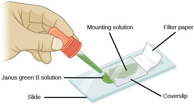
Stain with Janus Green B by using the wicking method as shown in Figure 8.1. Place a piece of filter paper or tissue on one side of the cover slip. Add one or two drops of Janus Green on the opposite site of the coverslip close to the edge. The stain solution will flow in by capillary action.
Monitor the changes in stain appearance for five to 10 minutes, taking turns observing the slide. Record your observation in your lab notebook.
Step 4: Critical Analysis: Draw and label all the structures that you can identify. Do not forget to add a title and the final magnification to all the drawings. Draw only what you observe. Do not copy from existing micrographs from published or online work. Compare the effect of different mounting solutions to distilled water. Did you observe a change in the color of mitochondria? Did it change over time? If there was an effect, how can you explain your observations? Can you think of other experiments that would support your conclusions?
Guided Inquiry
Step 1: Hypothesize/Predict: Janus Green B is a vital stain and an indicator of redox state. Knowing that it appears blue/green in its oxidized state and loses its color when it is reduced, allows you to predict which organelle will show a progressive change in color because it is the active site of oxidation-reduction and which organelle will appear colorless. What environmental conditions would be essential to observe a blue color of stain?
Step 2: Student-led planning: Prepare a wet mount of tissue under the microscope. Stain with Janus Green B, using the wicking method described earlier, while observing under the microscope. Which organelles can you distinguish? Are there any changes with time? Record your observations in your notebook. Repeat your experiment with a different mounting medium, staining with Janus Green B according to the wicking method. Record your observations in your notebook.
Step 3: Critical analysis: How did the various mounting fluids you used influence the response of the mitochondria to Janus Green B? How can you explain the effect that you observed? Compare the effect of different mounting solutions to distilled water. Was there an effect? If there was an effect, how can you explain your observations? Can you think of other experiments that would support your conclusions? Write your ideas in your notebook.
Assessments
- What do you predict would be observed if the epidermal layer of an onion is incubated in a solution of rotenone, an inhibitor of respiration?
- A student carefully mounts a specimen of onion epidermal layer, pushing out all the air bubbles. She is very disappointed that she does not observe a change in the color of Janus Green B. Can you explain her observation?
- Cyanide is a known metabolic poison that acts mainly by blocking cytochrome oxidase, an enzyme embedded in the inner membrane of mitochondria, and preventing the reduction of oxygen. If cyanide were added to an onion layer stained with Janus Green B, what you would observe and why?
