Central Vacuole and Anthocyanins
Learning Objectives
After completing the lab, the student will be able to:
- Make wet mounts of red onion skin.
- Describe the effect of pH on red onion skin.
Activity 2: Pre-Assessment
- How do plants and animals capture energy that they use to survive?
- Describe aspects of the morphological (physical structure) and behavioral characteristics of plants and animals that reflect their different means of acquiring energy.
- Discuss the answers to questions 1 and 2 with a partner and then the class.
Activity 2: Central Vacuole and Anthocyanins
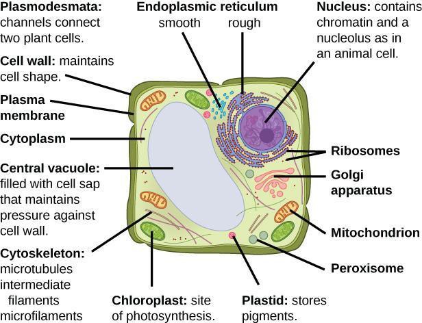
Plant cells are eukaryotic; they have subcellular organelles. Like bacteria, they have a cell wall to help keep the cell rigid—although in plants the cell wall is composed of a complex carbohydrate called cellulose. Plant cells also have a nucleus with DNA and a central vacuole (see Figure 5.6) full of water and other important substances (such as carbohydrates, non-nutrients, wastes) for maintaining life and to help maintain cell pressure.
The second major type of pigment found in plastids is water soluble. These pigment molecules, called flavonoids, are stored in the central vacuole of plants. The specific pigment found in the central vacuole that is examined in this activity is called anthocyanin (meaning flower blue); the addition of a sugar group to its structure makes it water soluble. If cations (positively charged ions or polyatomic ions) are added or removed from the anthocyanin structure the pigment color will change, showing the anthocyanins are sensitive to pH changes. Anthocyanins are found in most plant tissues as well as algae and some bacteria. Anthocyanins are hypothesized to protect plant tissues from harmful UV radiation and are also used as camouflage from herbivores.
Safety Precautions
- Be careful handling glass slides; the edges may be sharp.
- Observe proper use of the microscope; avoid handling the electric cord with wet hands.
- Do not use the coarse adjustment knob of the microscope at higher magnifications.
- Inform your teacher immediately of any broken glassware, as it could cause injuries.
- Wear goggles when handling pH solutions.
- If any pH solutions get on your hands, flush with water to remove.
- Dispose of pH solutions according to teacher instructions and local regulations.
- Wash your hands with soap and water after handling pH solutions.
For this activity, you will need the following:
- Red onion
- Clean microscope slides and cover slips
- Water and dropper
- Forceps
- pH solutions in dropper bottles (pH 3.0, pH 7.0, and pH 8.5)
- Notebook to observe and draw features of plant cells. For this activity, you will work in pairs.
Structured Inquiry
Step 1: Hypothesize/Predict: What subcellular features do you expect to see in red onion cells? Will the central vacuole, nucleus, and plastids (including chromoplasts) be obvious? What do you hypothesize will happen to the red onion cells in various pH solutions? Record your ideas and hypothesis in your notebook.
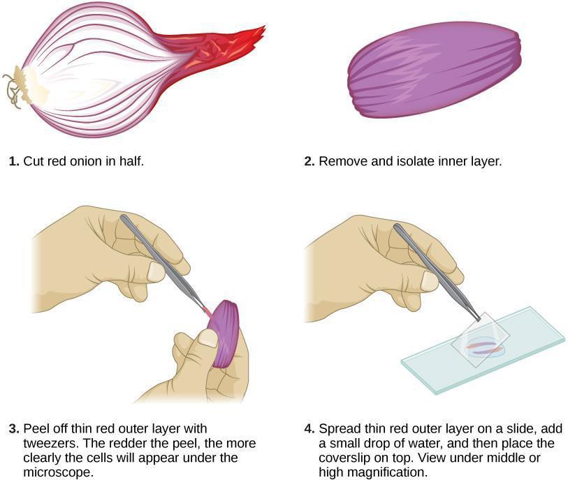
Step 2: Student-led planning: Prepare your plant cell slides as shown in Figure 5.7. Each partner is expected to prepare one sample. Each of you should view, draw, state the size and magnification, and label each sample. To prepare the onion skin, cut the onion in quarters, take the outer red peel of an onion section, and place it on the microscope slide.
Experiment with a few different cuts to determine the best technique to get the thinnest possible slice. Spread the onion skin on the microscope slide, put one small drop of water, and top with a coverslip. Record your observations (drawings, color if present, labels, magnification, and size of cell) in your notebook, keeping in mind your hypotheses from above.
Step 3: Critical Analysis: Were the predictions you made about what subcellular structures would be visible in an unstained specimen supported by your data (observations)? Why or why not? Are there any methods you could have used that would improve your results? Discuss with your partner and then write your answers in your notebook.
Guided Inquiry
Step 1: Hypothesize/Predict: A change in pH can often alter the function of macromolecules and cell structures. How do you think a change in pH will affect the pigment of the red onion cells? Write one hypothesis each describing how an increase and a decrease in pH would affect the onion cell pigment.
Step 2: Student-led planning. Your teacher has provided you with solutions of different pH. Discuss, with your partner, how you will examine each onion skin under the different pH solutions (e.g., How long should you observe each slide? How many times should you repeat your observations?). Record your plan in your notebook and create date tables in which to record your results.
Step 3: Carry out the experiment you designed in Step 2, preparing your slides using the technique in Figure 5.7 Create drawings in your notebook to record all of your observations.
Step 4: Critical analysis: Did the results you predicted match what you observed as the pH changed? In a paragraph, describe how increasing and decreasing pH affected the red onion cells. Include an explanation of why you think pH affected the cells in the way you observed. Discuss your analysis with your partner and write it in your notebook.
Assessments
- How were you able to find the vacuole of the red onion cells? What should you be looking for in the cell? Could you see individual chromoplasts?
- Red wines range in color from pink, red, and even violet-blue. If red grapes are used to make red wine, what part of the grape would explain that coloration? Predict the pH of wine based on the color.
