Meiosis and Fertilization
Learning Objectives
After completing the lab, the student will be able to:
- Read a karyotype.
- Determine karyotype abnormalities and identify an associated disorder or syndrome.
Activity 3: Pre-Assessment
- Looking at Figures 13.3 and 13.6, compare the outcomes of mitosis versus meiosis.
- After meiosis, some daughter cells may not contain the correct number of chromosomes. What failure in the meiosis process could cause this to occur?
- Discuss the answers to questions with a partner (think, pair, share) and then the class.
Activity 3: Meiosis and Fertilization
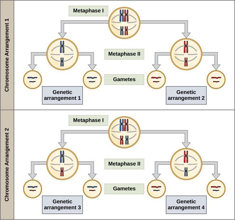
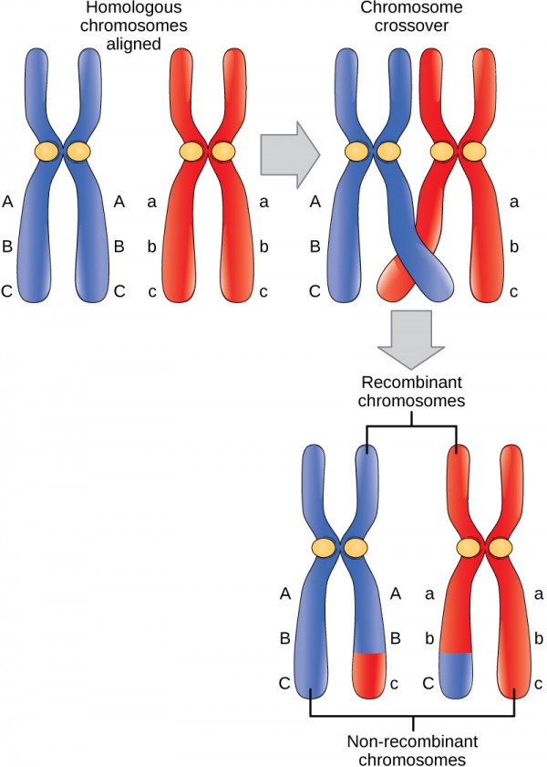
Meiosis is the process in sexually reproducing eukaryotes that forms sex cells or gametes, which include sperm and eggs (ova). To avoid doubling the number of chromosomes in each generation, reduction division (halving the number of chromosomes) in gamete production is necessary. Chromosomes are typically diploid (2N) or occur as double sets (homologous pairs) in each nucleus. Each homolog of a pair has the same sites or loci for the same genes. You might recognize that you have one set of chromosomes from your mother and the remaining set from your father. Meiosis reduces the number of chromosomes to a haploid (1N) or single set. Meiosis uses very similar mechanisms to mitosis. There are, however, several significant differences. The source cells for meiosis are found in the reproductive organs of animals (gonads: ovaries; oogenesis, where the eggs are produced and testes; spermatogenesis, where the sperms are produced) and plants (flowers: ovaries and anthers). Major differences between mitosis and meiosis include the association of homologous chromosomes (sister chromatids of the same chromosomes) attached as a tetrad group of four, as well as crossover of chromosome regions during prophase I of meiosis I. Meiosis shuffles the genetic material so that each resulting cell carries a new and unique set of genes in a process of independent assortment. Crossover provides one source of genetic variation in offspring.
This video demonstrates the sperm producing cells of a male crane fly, a species with only 8 chromosomes per cell during meiotic division.
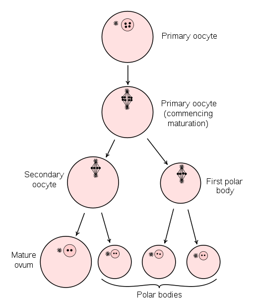
Oogenesis: In the human female genitalia reproductive structures, a growth process in which the primary egg cell (or ovum) matures into an ovum (1N)
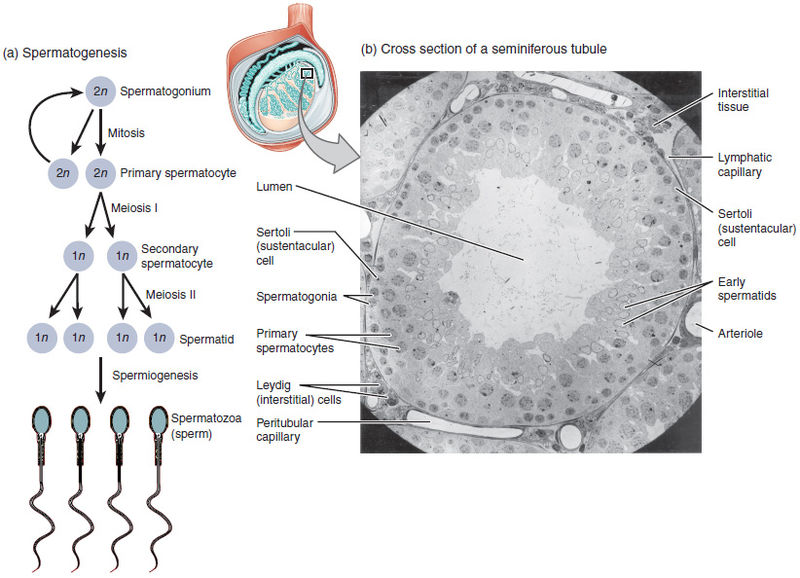
Spermatogenesis: In this process, haploid spermatozoa (1N) develop from germ cells in the seminiferous tubules of the testis male genitalia reproductive structures
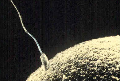
Fertilization: The union or fusion of the 2 gametes, ovum (1N) haploid and spermatozoa (1N) haploid in which the fertilized egg or zygote (2N) diploid has the double chromosome set restored. The size of the egg is emphasized by the sperm in contact
Humans have 23 different chromosomes. As stated, we have a diploid (2N) set of chromosomes: a full set of 23 chromosomes received from the sperm and another set of 23 from the ovum (egg) during fertilization. See Figure 13.7. During metaphase I of meiosis I, homologous chromosomes are separated. The chromosome number is reduced from diploid (2N) to haploid (1N) by the end of meiosis I, with each cell retaining duplicate sister chromatids. Meiosis II results in four haploid (1N) cells in sperm, each with only one set of each of the 23 chromosomes. In female humans, meiosis results in uneven division of the cytoplasm and the formation of two polar bodies (nuclei only) that are not involved in fertilization. One note about human fertilization: meiosis II is completed only after the sperm has entered the ovum.
There are several malfunctions that can occur during meiosis. Nondisjunction occurs when the sister chromatids in tetrads separate unevenly. Nondisjunction results in one cell getting an extra chromosome while another cell is missing a chromosome. Nondisjunction is associated with certain human genetic disorders. For example, Down syndrome is usually caused by nondisjunction that results in three copies of chromosome 21. Translocations can occur when chromosomes exchange genetic information with nonhomologous chromosomes. For a more detailed list, see this web page: https://www.genome.gov/11508982/chromosome-abnormalities-fact-sheet/
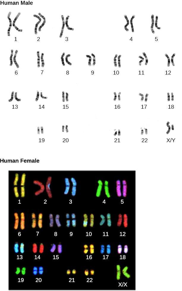
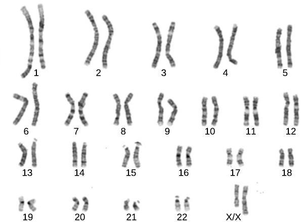
Karyotypes (chromosome spreads) are made by stopping cells in mitosis with a chemical and then dyeing with Giemsa stain. A picture is taken through a microscope and then digitally enlarged to see the chromosomal banding, or G-bands. Dark and light banding patterns help identify chromosomes and alterations to normal chromosomes. The chromosome spreads can be seen in Figure 13.7. Human females have two X chromosomes, and males have one X and one Y.
Safety Precautions
None
For this activity, you will need the following:
- Images of karyotypes
For this activity, you will work in pairs.
Structured Inquiry
Step 1: Hypothesize/Predict: A karyotype shows the number and appearance of chromosomes in the nucleus of a cell. Predict what you would look for in an abnormal karyotype. Record your predictions in your notebook.
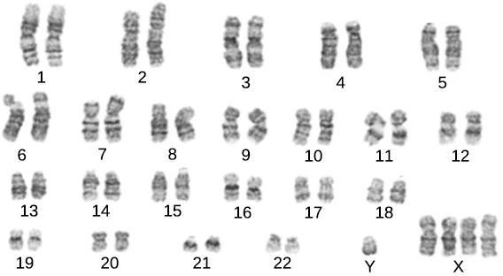
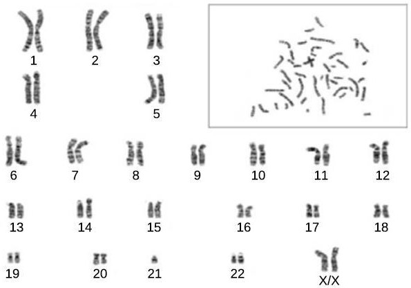
Step 2: Examine the three karyotypes 1, 2, and 3 shown in Figure 13.8, Figure 13.9, and Figure 13.10 respectively (below). Compare these karyotypes to the normal karyotypes shown in Figure 13.7 (above). Can you tell if the individual is female, male, or indeterminate (does not have a normal distribution of sex chromosomes)? Record any abnormalities in your notebook, and research the meaning of the changes in chromosome number or appearance.
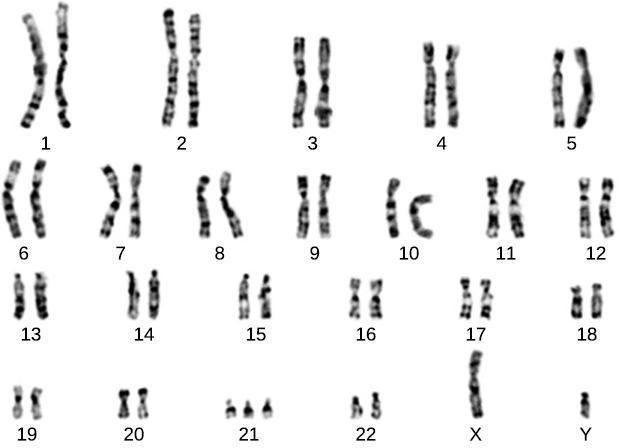
Step 3: Critical Analysis: What are the implications of nondisjunction in a karyotype? Using Karyotype 1 (Figure 13.8) as an example, explain the implications of the nondisjunction malfunction during meiosis for the chromosome count of the daughter cells.
Assessments
- This Robertsonian translocation involves a fusion of the long arms of two non-homologous chromosomes during meiosis. A patient possesses a Robertsonian translocation among chromosome 14 onto chromosome 13. No genetic material is missing from either chromosome. Explain why it is possible that there is no abnormality in the person but could be in the patient’s offspring.
Chromosomes - Crossing over of chromosomes during prophase I is common. How does this process increase the genetic diversity of the daughter cells?
