Comparing Plant Cells
Learning Objectives
After completing the lab, the student will be able to:
- Make wet mounts of bacteria, plant, and animal cells and view them under the microscope.
- Observe and identify differences between cells and cell structures under low and high magnification and record your observations.
- Explain how and why microscope stains are used when viewing cells under the microscope.
Activity 2: Pre-Assessment
- What new structures would you observe in Elodea cells that are not present in a cyanobacterium cell?
- Which of those structures would you expect to observe in an onion skin cell? Can you explain why some structures will be present in an Elodea cell but not in an onion epidermal cell?
- Discuss the answers to questions 1 and 2 with a partner and with the class.
Activity 2: Comparing Plant Cells
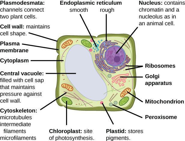
Plant cells are eukaryotic; they are larger than prokaryotic cells and have subcellular organelles. Like bacteria, they have a cell wall to help keep the cell rigid—in plants, the cell wall is composed of a complex carbohydrate called cellulose. Plant cells, like that shown in Figure 4.4, also have a nucleus with DNA and a large central vacuole full of water and other important substances for maintaining life, such as carbohydrates, non-nutrients, and wastes, and help maintain cell pressure. Many plant cells also have chloroplasts with chlorophyll where photosynthesis occurs and/or other membrane-bound plastids, such as amyloplasts that store starch or chromoplasts that store pigments.
Safety Precautions
- Be careful when handling glass slides; the edges may be sharp.
- Dispose of used cover slips in a glass disposal box.
- Observe proper use of the microscope; avoid handling the electric cord with wet hands.
- Do not use the coarse adjustment knob of the microscope at high and oil immersion objectives.
- Inform your teacher immediately of any broken glassware, as it could cause injuries.
- Wash your hands with soap and water after handling live organisms.
For this activity, you will need the following:
- Elodea anacharis, moss, or Spirogyra green algae
- Light compound microscope
- Clean microscope slides and cover slips
- Lens paper
- Water and dropper
- Yellow onion
- Potato
- Forceps
- Iodine solution
For this activity, you will work in pairs.
Guided Inquiry
Step 1: Hypothesize/Predict: What features do you expect to see in the plant cell Elodea and the onion skin under low and high magnification? Draw and label your prediction in your notebook.
Step 2: Student-led Planning: Prepare a wet mount of Elodea, moss, or Spirogyra by removing a small piece with forceps, placing it on the middle of the slide, and adding a small drop of water. Place one edge of the coverslip in the water and drop onto the specimen to remove air bubbles. To prepare a wet mount of onion, remove a thin piece of the onion skin with forceps, add one drop of iodine solution to act as a stain, and place a coverslip on top of the specimen. To prepare a wet mount of potato, scrape potato cells onto the slide, add iodine and a coverslip. Record your observations as drawings. Use color if present, label the magnification, and estimate the size of the cells in your notebook. Each partner is expected to prepare one sample. Each person should view, draw, state the size and magnification, and label each sample.
Step 3: Critical Analysis: What differences are you expecting to see between Elodea, onion, and potato? What similarities? If you used iodine as a stain, did that reveal any other differences in the plants? Why would these organisms have anything in common based on your prediction in Step 1?
Assessments
- Were the features you predicted to see in Elodea cells and the onion cell visible at low and high magnification? Which structures in Figure 4.4 can you identify in the Elodea cells? What about the onion cells? Why do you think this is the case?
- What are the similarities and differences between a cyanobacterium and a plant cell?
