Observation of Anabaena or Nostoc
Learning Objectives
After completing the lab, the student will be able to:
- Make wet mounts of bacteria, plant, and animal cells and view them under the microscope.
- Observe and identify differences between cells and cell structures under low and high magnification and record your observations.
Activity 1: Pre-Assessment
- What is the difference between the cells of a bacterium and the cells of your own body?
- Compare and contrast the structures of prokaryotic and eukaryotic cells.
- Discuss the answers to questions 1 and 2 with a partner and then the class.
Activity 1: Observation of Anabaena or Nostoc
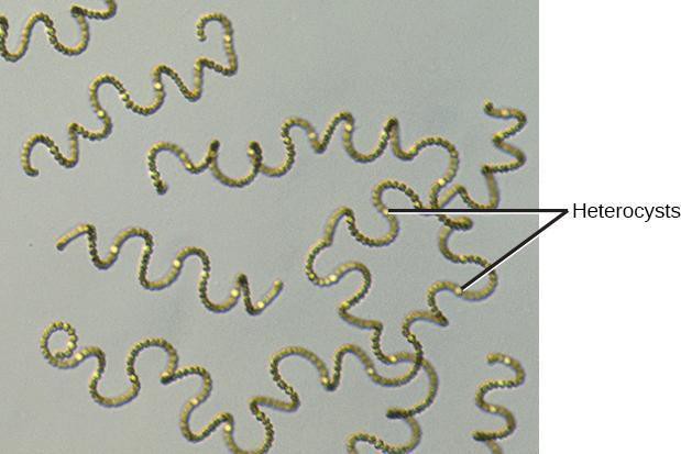
Prokaryotes, unicellular organisms lacking a nucleus, include cyanobacteria (formerly blue-green algae. This name is now considered inaccurate because algae are eukaryotes). Cyanobacteria, like those shown in Figure 4.1, are photoautotrophs—organisms that carry out photosynthesis by using light energy, water, and carbon dioxide from the air and converting to sugars, and providing oxygen to the atmosphere as a waste product. Cyanobacteria contain pigments capable of capturing light energy but do not contain chloroplasts. Cyanobacteria are single-celled organisms, but some can form colonies with differentiated cell types. For example, some species can form specialized cells called heterocysts—structures containing enzymes that can take atmospheric nitrogen (nitrogen fixation) from the air and convert it into usable molecules for DNA, RNA, and protein synthesis. Nitrogen fixation involves converting nitrogen gas (N2) from the atmosphere to ammonia (NH3). Ammonia is a form of nitrogen that can be used to build other molecules, including DNA, RNA, and proteins. Oxygen, a waste product of photosynthesis, interferes with a key enzyme in nitrogen fixation. Thus, only a few cells in a colony (about 1 in 10) become heterocysts. Resources, such as ammonia and sugars from photosynthesis, are then shared between cells. These cells range from 1–40 micrometers in size. Not all bacteria can carry out photosynthesis. The majority of bacteria are heterotrophic and they live virtually everywhere on Earth. These cells are much smaller, ranging from 0.5–8 micrometers. Eukaryotic cells have many more features and organelles and range in size from 10–500 micrometers.
Safety Precautions
- Be careful when handling glass slides; the edges may be sharp.
- Observe proper use of the microscope; avoid handling the electric cord with wet hands.
- Do not use the coarse adjustment knob of the microscope when using the high and oil immersion objective
- Inform your teacher immediately of any broken glassware, as it could cause injuries.
- Wash your hands with soap and water after handling live organisms.
For this activity, you will work in pairs.
For this activity, you will need the following:
- Light compound microscope (10×, 40×, 100×)
- Lens paper
- Prepared slide of Anabaena or Nostoc or images of Anabaena or Nostoc
- Special slide with micron ruler or clear millimeter ruler (you can photocopy ruler on overhead transparency, then cut and tape to microscope slide)
Structured Inquiry
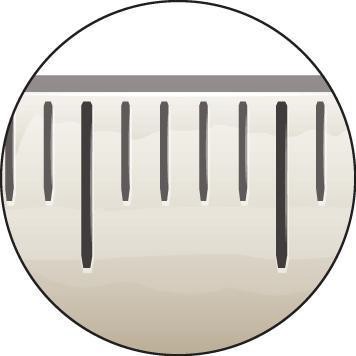
Step 1: Estimate the size of the field of view at all the magnifications of your microscope by placing a clear millimeter ruler on the stage of the microscope. This will allow you to estimate the sizes of cells. Convert your millimeter estimates to micrometers for this activity.
Step 2: Hypothesize/Predict: In your notebook predict (draw) what you would expect to see in the microscope. How big do you predict the cells will be? What features do you expect to see? Do you expect to see organelles or a cell wall?
Step 3: In your notebook, create a detailed drawing, with a sharp pencil, of the structure of the cyanobacterium. An example of a detailed drawing is seen in Figure 4.3. Record the estimated size of the cells at the magnification used. Use color in your drawings if appropriate. Identify the colors used and label any obvious structures. Note the shapes or organization of the cells.
Step 4: Critical Analysis: Think about the cell types you observed. Do your observations match your expectations? Given that the cyanobacteria are photosynthetic, was the color what you expected? Why? Did you expect the cells to have organelles? Did they? Did they have a cell wall? Did you find heterocysts? Discuss with your partner and write your answers in your notebook.
Assessments
- Create a diagram of a general prokaryotic cell and a general eukaryotic cell. Label the cell structures that differ between the two cell types.
- How would internal membrane-bound structures, such as chloroplasts and mitochondria, allow chemical reactions to occur more efficiently in cells?
