Observation of Subcellular Structures in Animal Tissues
Learning Objectives
After completing the lab, the student will be able to:
- Observe Nissl bodies in a neuron as an example of endoplasmic reticulum.
- Observe striated muscle as an example of cytoskeleton thin filaments.
- Visualize mitochondria using a biological stain.
Activity 3: Pre-Assessment
- What structures would you expect to find in plant cells but not animal cells?
- Do you expect that the subcellular structures will be easy to see? Why or why not?
- Discuss the answers to questions 1 and 2 with a partner (think, pair, and share) and then the class.
Activity 3: Observation of Subcellular Structures in Animal Tissues
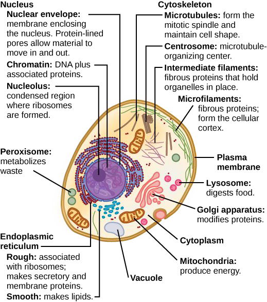
Animal cells (Figure 5.8) are eukaryotic and possess subcellular components in common with the plant cells you observed earlier. Some examples include nuclei (contains DNA, controls cell function), Golgi apparatus (sorting, packing, and modification of proteins), mitochondria (energy production from organic molecules such as glucose), ribosomes (translation of messenger RNA into proteins), and the endoplasmic reticulum (folding of proteins and manufacture of lipids). Animal cells are predominantly colorless. Exceptions include the red of hemoglobin in red blood cells, red myoglobin in striated muscle, and melanin in skin cells. Thus, specific biological stains are required to make visible cellular features under a light microscope. Janus Green B stain is effective for viewing mitochondria, chromosomes, and the rough endoplasmic reticulum.
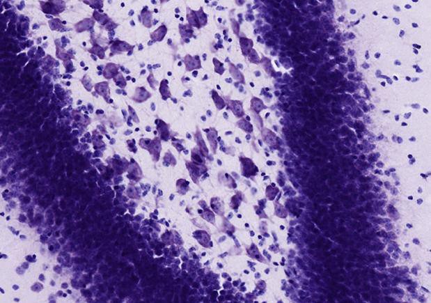
Nissl bodies, shown in Figure 5.9, present in neurons can be stained and allow you to see the rough endoplasmic reticulum and rosettes of free ribosomes. The cytoskeleton includes thin and thick filaments, which are involved in cell shape, movement, and muscle contractions.
Safety Precautions
- Be careful handling glass slides; the edges may be sharp.
- Observe proper use of the microscope; avoid handling the electric cord with wet hands.
- Do not use the coarse adjustment knob of the microscope at higher magnifications.
- Inform your teacher immediately of any broken glassware, as it could cause injuries.
- Wear goggles when using staining solutions.
- Dispose of staining solutions according to teacher instructions and local regulations.
- Wash your hands with soap and water after handling live organisms and biological stains.
For this activity, you will need the following:
- Prepared slides of Nissl bodies and striated muscle
- Elodea anacharis
- Clean microscope slides, cover slips
- Janus Green B stain
- Dropper
- Small squares of paper towels
- Notebook to observe and record data. For this activity, you will work in pairs.
Structured Inquiry
Step 1: Hypothesize/Predict: Predict how big the structures of the Nissl bodies, thin and thick filaments, nuclei, and mitochondria will appear differently when you look at them under the microscope. Which structures do you think you can see without staining?
Step 2: Observe the prepared slides and record (draw, label any visible parts, use color if visible, record magnification and size of cells) in your notebook.
Step 3: Critical analysis: How visible were the Nissl bodies, thin and thick filaments, and mitochondria before and after staining? Write your answers in your notebook.
Guided Inquiry
Step 1: Hypothesize/Predict: Janus Green B stains more when more oxygen is present. Given the function of mitochondria, predict how a biological stain would help you visualize mitochondria. Record your prediction in your notebook.
Step 2: Prepare a wet mount microscope slide with Elodea anacharis. Experiment with a few different cuts to determine the best technique to get the thinnest possible slice. Observe your prepared slide, record (draw, label any visible parts, use color if visible, record magnification and size of cells) in your notebook. Each partner should prepare a slide and observe and report on both slides.
Step 3: Each student should prepare a second wet mount, this time with Janus Green B stain, as in Figure 5.9. Observe over a period of time. Record your observations, draw, label any visible parts, use color if applicable, and record magnification in your notebook.
Step 4: Critical Analysis: Were the structures smaller or larger than you thought? There should be small blue dots visible on the Elodea cells—much smaller than the chloroplasts. Hypothesize what those blue dots might be. Record the answers to these questions in your notebook.
Assessments
- Write a description that would allow another student to distinguish stained mitochondria, nuclei, and features of the rough endoplasmic reticulum from each other through a microscope.
