Extracting Pigments from Plant Material
Learning Objectives
After completing the lab, the student will be able to:
- Extract pigments from plant material.
- Separate pigments by paper chromatography.
Activity 1: Pre-Assessment
- Think about what colors you have seen in leaves of plants. What determines the color of a leaf? What color or colors do you expect to see if you extract pigments from leaves and separate them chemically?
- Other structures in plants besides leaves are brightly colored. Fruit, flowers, and some roots display colors. Are these colors associated with photosynthesis? What are the purposes of those colors?
- Discuss the answers to questions 1 and 2 with the class.
Activity 1: Extracting Pigments from Plant Material
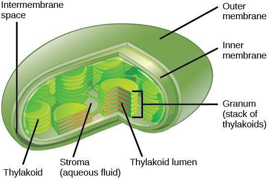
The chloroplast, as illustrated in Figure 10.1, is the photosynthetic organelle in the plant cell. The chloroplast is surrounded by a double membrane. Within the inner membrane is a fluid compartment called the stroma. The thylakoids form a complex network of membranes that appear as stacked disks called grana (singular, granum). The photosystems, and photosynthetic pigments they contain, are embedded in the thylakoid membrane. Carbon fixation takes place in the stroma. What chemical properties would you expect photosynthetic pigments to display and how can these chemical properties be used to extract the pigments from tissues? How do the solubility properties enable scientists to separate individual pigments for analysis?
Safety Precautions
- Wash your hands after completion of this activity.
For this activity, you will need the following:
- Plant material: intact leaves of spinach and Coleus (one leaf of each per pair of students)
- Filter or chromatography paper
- Ruler (one per group)
- Coins
- Pencils
- Colored pencils
- Forceps
- Scissors
In this activity, you will work in pairs.
Structured Inquiry
Step 1: Hypothesize/Predict: In this activity, you will extract pigments from spinach leaves. Discuss and predict what colored pigments you will observe after extraction. Write your hypotheses in your notebook.
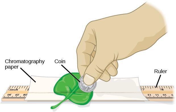
Step 3: Complete the steps below to extract pigments from plant material, as illustrated in Figure 10.2. Use the method you devised in step 2 to avoid touching the paper with your fingers.
- Measure the height and the diameter (D) of the container with your partner to determine the size of the piece of chromatography paper you need for the experiment. Subtract 1 cm from the height of the container. The width of the chromatography paper should be about 1 cm shorter than the circumference of the container. Record your calculations and show them to your instructor before you cut the paper.
- With a pencil, draw a line across the entire width of filter/chromatography paper about 1.5 cm from the bottom. This line is called the origin or start of the separation. It is the place where the sample is applied.
- Select 1 large spinach leaf and carefully blot it dry with paper towels. Note in your lab notebook why it is important to blot dry the leaf.
- Position the leaf on top of the pencil line leaving a space on each end. The leaf should reach the edge of the paper.
- Align a plastic ruler over the leaf with the edges of the pencil line that was left uncovered by the leaf.
- Roll a coin pressing down firmly along the ruler edge without crushing the rest of the leaf and smearing pigment on the surface of the filter paper. You may find it helpful to lay the filter paper on top of the paper towels taking care not to cover the line where the pigments are deposited.
- Let the pigment line dry for 3 to 5 minutes. Move to a different section of the leaf and roll the coin again over the same line. Keep the band of pigments as narrow as possible. The pigments will diffuse during separation and the bands will smear if they are laid down too thick.
Step 4: Critical Analysis: Record the results from your experiment. Try to identify the pigment or pigments you extracted. Can you state conclusively what types of pigments are present in your extract on the line? How could you determine which pigments are present in the extract without performing a chemical separation? Write your answers in your lab notebook. You may refer to Figure 10.2 while discussing the answer.
Guided Inquiry
Step 1: Hypothesize/Predict: If pigments are colored molecules, can you decide which pigments are present by looking at biological material? Do spinach leaves contain other pigments besides the green chlorophylls? Do Coleus leaves contain additional pigments? Do you see a red pigment on the line for Coleus? Write your hypotheses in your notebook.
Step 3: Critical Analysis: Observe the pigment lines on the paper. Record in your lab notebook the color or colors you detected. Note the differences, if any, between the spinach and Coleus samples.
Assessments
- Plants in the rainforest that live under the canopy of tall trees must adapt to low light conditions. Can you predict how the pigments of plants that receive low light levels will differ from the pigments of plants that receive high light levels? What would happen to the forest if the tall trees were removed for timber?
- You analyze a pigment extract from a mutant plant and discover that the plant defective in its carotenoid biosynthetic pathway. What would happen to that plant if it is grown in bright sunlight? Where would you expect it to thrive and how would such a plant compensate for the loss of carotenoids?
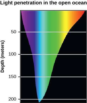
- The Rhodophyta are algae that can grow deeper in the ocean than other plants. Based on the information shown in Figure 10.3, what color of the visible spectrum would you expect to be absorbed by an accessory pigment found in Rhodophyta? Why do you think plants have reddish colors?