Audition
Anatomy and Physiology of the Ear
Watch this video:
Media 17.2 Hearing & Balance: Crash Course A&P #17 [Online video]. Copyright 2015 by CrashCourse.
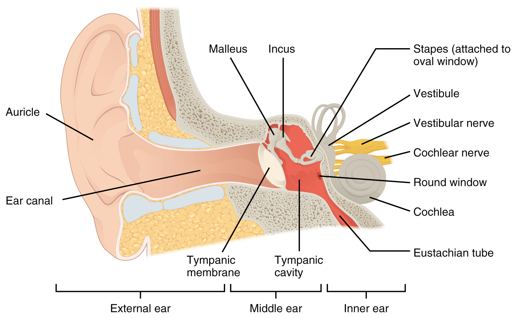
Hearing, or audition, is the transduction of sound waves into a neural signal that is made possible by the structures of the ear (see Figure 17.3).
Structure of the Ear
The structure of the ear can be divided into three parts:
The external ear consists of the auricle, sometimes referred to as the pinna, and the ear canal. The C-shaped curves of the auricle direct sound waves toward the auditory canal. This canal enters the skull through the external auditory meatus of the temporal bone. At the end of the auditory canal is the tympanic membrane, a structure that vibrates when struck by sound waves and separates the ear canal from the middle ear space.
The middle ear consists of the ossicles, the oval window, and the Eustachian tube. The three ossicles are the malleus, incus, and stapes, which are Latin names that roughly translate to hammer, anvil, and stirrup. The malleus is attached to the tympanic membrane on one end and articulates with the incus on the other. The incus, in turn, articulates with the stapes. The stapes is then attached to the inner ear, where the sound waves are transduced into a neural signal. Vibrations of the ossicles travel through the oval window, moving fluid in a wave-like motion. The frequencies of the fluid waves match the frequencies of the sound waves. The middle ear is connected to the pharynx through the Eustachian tube, which helps equilibrate air pressure across the tympanic membrane. The tube is normally closed but will pop open when the muscles of the pharynx contract during swallowing or yawning.
The inner ear is often described as a bony labyrinth, as it is composed of a series of canals embedded within the temporal bone. It consists of the cochlea, which is responsible for hearing, and the vestibule, which is responsible for balance. The neural signals from these two regions are relayed to the brain stem through separate fiber bundles. However, these two distinct bundles travel together from the inner ear to the brain stem as the vestibulocochlear nerve. Sound is transduced into neural signals within the cochlear region of the inner ear, which contains the sensory neurons of the spiral ganglia. These ganglia are located within the spiral-shaped cochlea of the inner ear.
Image Descriptions
Figure 17.3 image description: This image shows the structure of the ear with the major parts labeled. The ear is divided up into 3 parts from left to right: external ear, middle ear, and inner ear. Labels for each part read: external ear (auricle, ear canal), middle ear (tympanic membrane, malleus, incus, tympanic cavity), inner ear (stapes, vestibule, vestibular nerve, cochlear nerve, cochlea, round window, eustachian tube). [Return to Figure 17.3].
