Anatomy and Physiology of the Male Reproductive System
Anatomy of the Male Reproductive System
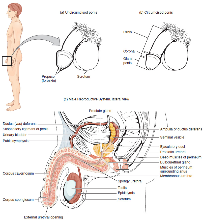
The anatomy of the male reproductive system consists of structures that include the testes, the epididymis, the penis, and the ducts and glands that produce and carry semen. Sperm exit the scrotum through the vas deferens. The spermatic cord is an enclosed sheath that includes the vas deferens, arteries, veins, and nerves. The seminal vesicles and prostate gland add fluids to the sperm to create semen.
Physiology of the Male Reproductive System
The anatomy of the male reproductive system consists of structures that include spermatogenesis, sperm, sperm transport, epididymis, ducts, prostate gland, and bulbourethral glands.
Spermatogenesis
Spermatogenesis occurs in the seminiferous tubules that form the bulk of each testis. The process begins at puberty, after which time sperm are produced constantly throughout a man’s life. One production cycle takes approximately 64 days. One production cycle is considered from spermatogonia through to formed sperm. A new cycle starts approximately every 16 days, although this timing is not synchronous across the seminiferous tubules.
Sperm
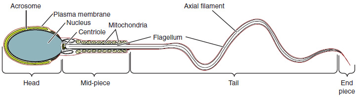
Sperm are smaller than most cells in the body; in fact, the volume of a sperm cell is 85,000 times less than that of the female gamete. Approximately 100 to 300 million sperm are produced each day, whereas women typically ovulate only one oocyte per month. As is true for most cells in the body, the structure of sperm cells speaks to their function. Sperm have a distinctive head, mid-piece, and tail region (see Figure 14.2).
Sperm Transport
To fertilize an egg, sperm must be moved from the seminiferous tubules in the testes, through the epididymis, and—later during ejaculation—along the length of the penis and out into the female reproductive tract. It takes an average of 12 days for sperm to move through the coils of the epididymis, with the shortest recorded transit time in humans being one day.
Epididymis
Sperm enter the head of the epididymis and are moved by the contraction of smooth muscles lining the epididymal tubes. As the sperm mature, they acquire the ability to move under their own power. Once inside the female reproductive tract, they will use this ability to move independently toward the unfertilized egg. The more mature sperm are then stored in the tail of the epididymis until ejaculation occurs.
Ducts
During ejaculation, sperm exit the tail of the epididymis and are pushed by smooth muscle contraction to the vas deferens (also called the ductus deferens). The vas deferens is a thick, muscular tube that is bundled together inside the scrotum with connective tissue, blood vessels, and nerves into a structure called the spermatic cord. From each epididymis, each vas deferens extends through the inguinal canal in the abdominal wall and continues to a region called the ampulla. The sperm is mixed with fluid from the paired seminal vesicles and moves into its associated ejaculatory duct. The ejaculatory ducts transport the seminal fluid to the prostate gland.
Prostate Gland
The prostate gland secretes an alkaline, milky fluid to the passing seminal fluid (referred to as semen) to first coagulate and then decoagulate the semen following ejaculation. The temporary thickening of semen helps retain it within the female reproductive tract. Once decoagulated, the sperm can pass farther into the female reproductive tract.
Bulbourethral Glands
Bulbourethral glands release a thick, salty fluid that lubricates the end of the urethra and vagina and helps to clean urine residues from the penile urethra.
Image Descriptions
Figure 14.1 image description: This figure shows the different organs in the male reproductive system. The top panel shows the side view of a man and an uncircumcised and a circumcised penis. The bottom panel shows the lateral view of the male reproductive system and the major parts are labeled. [Return to Figure 14.1].
Figure 14.2 image description: This diagram shows the structure of sperm; the major parts are labeled (from left to right): head section (acrosome, plasma membrane, nucleus), mid-piece (centriole, mitochondria, flagellum), tail (flagellum, axial filament), end piece (end piece). [Return to Figure 14.2].
