Anatomy of the Blood Vessels
Anatomy of the Blood Vessels
Blood pumped by the heart flows through a series of vessels known as arteries, arterioles, capillaries, venules, and veins before returning to the heart.
- Arteries transport blood away from the heart and branch into smaller vessels, forming arterioles.
- Arterioles distribute blood to capillary beds, the sites of exchange with the body tissues.
- A capillary is a microscopic channel that supplies blood to the tissues themselves, a process called perfusion.
- Exchange of gases and other substances occurs in the capillaries between the blood and the surrounding cells and their tissue fluid (interstitial fluid).
- For capillaries to function, their walls must be leaky, allowing substances to pass through.
- Capillaries lead back to small vessels known as venules.
- Venules are small veins that converge into larger veins.
- A vein is a blood vessel that conducts blood toward the heart.
- Compared to arteries, veins are thin-walled vessels with large and irregular lumens.
- Larger veins are commonly equipped with valves that promote the unidirectional flow of blood toward the heart and prevent backflow toward the capillaries caused by the inherent low blood pressure in veins as well as the pull of gravity.
- Other ways in which the body assists the transport of venous blood back to the heart involve contractions of skeletal muscles in the extremities (see figure below) as well as pressure variations caused by the breathing motion of the chest.
Both arteries and veins have the same three distinct tissue layers, called tunics (after the garments first worn by ancient Romans). From the most interior layer to the outer, these tunics are the tunica intima, the tunica media, and the tunica externa (see Figure 9.1). The smooth muscle in the middle layer, the tunica media, provides the vessel with the ability to vasoconstrict and vasodilate as needed to ensure sufficient blood flow.
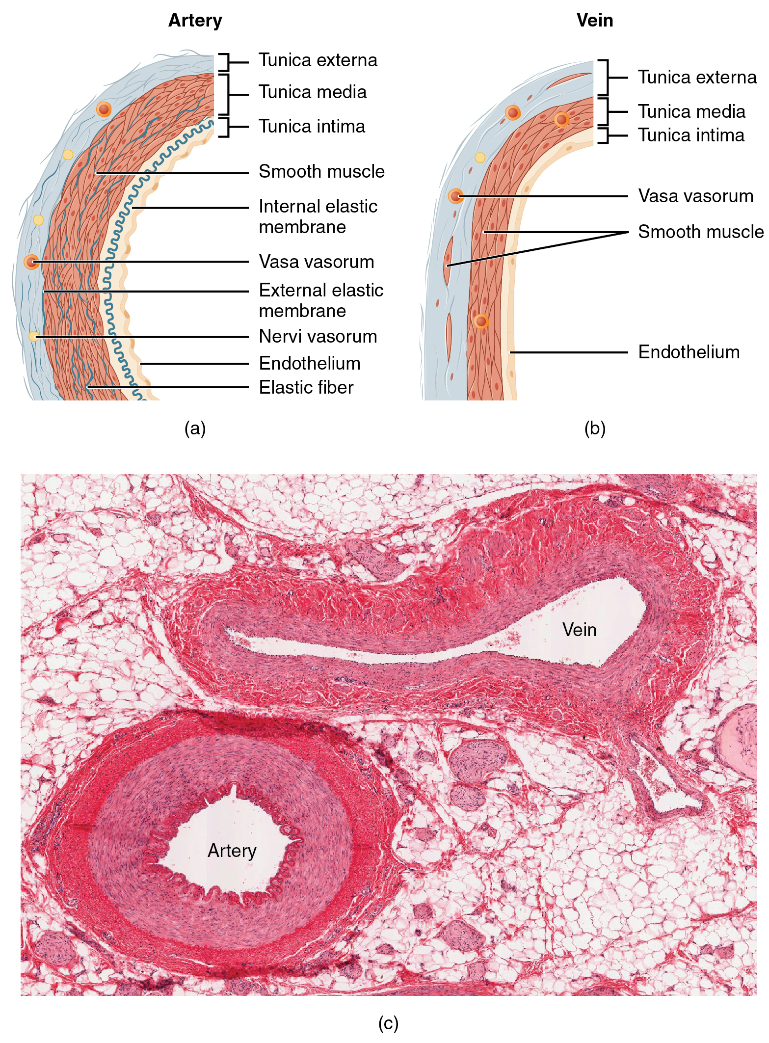
Image Descriptions
Figure 9.1 image description: The top left panel of this figure shows the ultrastructure of an artery (labels read from top: tunica externa, tunica media, tunica intima, smooth muscle, internal elastic membrane, vasa vasorum, external elastic membrane, nervi vasorum, endothelium, elastic fiber), and the top right panel shows the ultrastructure of a vein (labels read from top: tunica externa, tunica media, tunica intima, vasa vasorum, smooth muscle, endothelium). The bottom panel shows a micrograph with the cross sections of an artery and a vein. [Return to Figure 9.1].
