Medical, Surgical, & Viewing Terms and Abbreviations
Medical Careers & Professional Terminology
radiologic technologist (RT): Creates images of patients’ bodies using medical equipment. The images help doctors diagnose and treat diseases and injuries. Technologists assist physicians in procedures as well as prepare and administer contrast agents in order to better visualize the anatomy of interest. Rad Techs need to be licensed in many states in order to administer x-rays (x-radiation). Many schools offer degrees or certificates to achieve before taking your registry in order to become licensed.
specialized radiologic technologist: Radiologic technologists can specialize in Computed Tomography (CT); Magnetic Resonance Imaging (MRI); Bone Densitometry (D); Positron Emission Tomography (PET); Nuclear Medicine (NM); and Radiation Therapy (RT). These specialties could be completed while on the job or with an additional year of education depending on the area chosen. Technologists can become certified in multiple modalities by completing requirements and passing the registry for the specified modality of interest.
radiologist: A medical doctor that specializes in diagnosing and treating injuries and diseases using medical imaging procedures, or exams, from the Radiology Department. Procedures could include x-rays, Computed Tomography (CT), Magnetic Resonance Imaging (MRI), Nuclear Medicine (NM), Positron Emission Tomography (PET), and Ultrasound (Sonography). Radiologists complete at least 13 years of training, including medical school, a four-year residency, and most often, an additional one- or two-year fellowship of very specialized training, such as radiation oncology, pediatric radiology, or interventional radiology. They are certified by the American Board of Radiology, and they have exacting requirements for continuing medical education throughout their practicing years.
physical therapist assistant (PTA): Provides physical therapist services under the direction and supervision of a physical therapist. PTAs implement components of patient care, obtain data related to the treatments provided, and collaborate with the PT to modify care as necessary. They assist patients in recovery after injuries, postoperative care, and pain management through strategic exercises and therapeutic methods. PTAs have completed a minimal associate-level degree and passed a licensing exam.
physical therapist (PT): Physicians who diagnose and treat patients by prescribing exercises, hands-on care, and patient education. PTs are required to obtain a doctorate in physical therapy. This typically takes 3 years, and graduates must also pass a state licensure exam.
orthopedic surgeon: A medical doctor who completes an additional 5 years of specialized training in the prevention, diagnosis, treatment, and surgery of disorders and diseases related to the musculoskeletal systems.
neurologist: A medical doctor who completes an additional 5 years of specialized training in the prevention, diagnosis, and treatment of disorders and conditions related to the brain, spinal cord, nerves, and muscles.
kinesiologist: A regulated health care professional with a 4-year degree in kinesiology or a related discipline. Kinesiologists work in a variety of settings that assist people with pain management, injury prevention, and health promotion through biomechanics.
rheumatologist: A medical doctor who has additional training as an internist with a subspecialty in rheumatology. These doctors have special interests in autoimmune disorders and their impact on the musculoskeletal system.
doctor of chiropractic (DC)/chiropractor: A health care practitioner with 7 years of education, supervised internships, and national examinations. They are trained in prevention, assessment, and treatment of the spine, muscular system, and nervous system. Chiropractors focus on spinal adjustments, nutrition, and preventing injury without the use of pharmaceuticals or surgical procedures.
Imaging Techniques/Procedures for the Organ System Built from Word Parts
arthrography: radiographic imaging of a joint (with contrast media)
myelography: radiographic imaging of the spinal cord (with contrast media)
arthroscopy: visual examination of a joint
electromyogram: record of the (intrinsic) electrical activity in the (skeletal) muscle
Imaging Techniques/Procedures for the Organ System Not Built from Word Parts
dual x-ray absorptiometry (DXA): radiographic imaging, usually of the lumbar spine and hips, to measure bone loss and bone mineral density; the procedure utilizes low doses of radiation and is used in the diagnosis of osteoporosis and monitoring of treatment (also called dual-energy x-ray absorptiometry [DEXA], bone densitometry, and bone density test)
bone markers: blood and urine tests to determine the rate of bone turnover (resorption and formation); often used with DXA to diagnose and monitor treatment of osteoporosis and other bone disorders
muscle biopsy: removal of muscle tissue using a needle or small incision; used to assess musculoskeletal abnormalities involving weakness or pain such as muscular dystrophy, myasthenia gravis, and polymyositis
Diagnostic Imaging Procedures
bone densitometry: a method of determining the density of bone by radiographic techniques used to diagnose osteoporosis; dual x-ray absorptiometry (DXA) is commonly used for this test
bone scan: (nuclear medicine test) used to detect the presence of metastatic disease of the bone and to monitor degenerative bone disease
magnetic resonance: used to evaluate the bones and soft tissue of the shoulders, hips, elbows, knees, ankles, feet, and spinal cord for stenosis, spinal cord defects, and degenerative disk changes
radiography: (radiographic imaging) of the bones and joints is used to identify fractures or tumors, monitor healing, or identify abnormal structures.
single-photon emission computed tomography (SPECT): of the bone is an even more sensitive nuclear method for detecting bone abnormalities
Surgical Techniques/Procedures Used in the Organ System
arthrocentesis: surgical puncture to aspirate fluid from a joint
arthrodesis: surgical fixation of a joint (a.k.a. joint fusion)
arthroplasty: surgical repair of a joint
bursectomy: excision of a bursa
carpectomy: excision of a carpal bone
chondrectomy: excision of cartilage
chondroplasty: surgical repair of cartilage
costectomy: excision of a rib
cranioplasty: surgical repair of the skull
craniotomy: incision into the cranium (as for surgery of the brain)
discectomy: excision of an intervertebral disk (a portion of the herniated disk is removed to relieve pressure on nerve roots; uses a larger incision than microdiscectomy)
fasciotomy: incision into fascia (to relieve tension or pressure)
laminectomy: excision of a lamina (often performed to relieve pressure on the nerve roots in the lower spine caused by a herniated disk and other conditions)
maxillectomy: excision of the maxilla
meniscectomy: excision of a meniscus (performed for a torn cartilage)
microdiscectomy: small excision of an intervertebral disk (minimally invasive surgery to remove a portion of the herniated disk to relieve pressure on nerve roots)
myorrhaphy: suturing of a muscle
osteotomy: incision into the bone
phalangectomy: excision of a finger or toe bone
rachiotomy: incision into the vertebral column
spondylosyndesis: fusing together of the vertebrae (also called spinal fusion)
synovectomy: excision of the synovial membrane (of a joint)
tarsectomy: excision of (one or more) tarsal bones
tenomyoplasty: surgical repair of the tendon and muscle
tenorrhaphy: suturing of a tendon
vertebroplasty: surgical repair of a vertebra (usually performed for compression fractures due to osteoporosis)
Abbreviations Commonly Used with the Organ System
Disease and Disorders
CTS: carpal tunnel syndrome
Fx: fracture
HNP: herniated nucleus pulposus
MD: muscular dystrophy
MG: myasthenia gravis
OA: osteoarthritis
PM: polymyositis
RA: rheumatoid arthritis
RSI: repetitive strain injury
Diagnostic
DEXA: (spoken as a whole word): dual-energy x-ray absorptiometry
DXA: (spoken as a whole word): dual x-ray absorptiometry
EMG: electromyogram
Treatment
THA: total hip arthroplasty
TKA: total knee arthroplasty
Medical Specialties
DC: Doctor of Chiropractic
DO: Doctor of Osteopathy
Ortho: Orthopedics
Descriptive
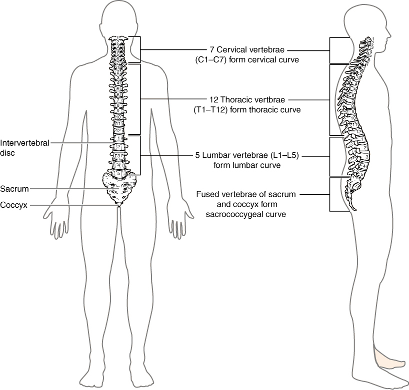
C1-C7: cervical vertebrae
T1-T12: thoracic vertebrae
L1-L5: lumbar vertebrae
Medical Terms Practice
For each card, click the speaker icon to hear the correct pronunciation of the listed term. Practice saying the term to yourself, then attempt to define the term from memory. Click “Turn” to flip the card and see the definition. Use the right and left arrows to toggle through the cards in each set.
Image Description
Figure 4.6 image description: This image shows the structure of the vertebral column. The left panel shows the front view of the vertebral column. Labels and the right panel show the side view of the vertebral column. labels read (from top): 7 cervical vertebrae (C1-C7) form cervical curve, 12 thoracic vertebrae (T1-T12) form thoracic curve, intervertebral disc, 5 lumbar vertebrae (L1-L5) form lumbar curve, Fused vertebrae of sacrum and coccyx form sacrococcygeal curve, sacrum, coccyx. [Return to Figure 4.6].
