Body Cavities, Serous Membranes, and Tissue Membranes
Body Cavities and Serous Membranes
The body maintains its internal organization by means of membranes, sheaths, and other structures that separate compartments. The dorsal (posterior) cavity and the ventral (anterior) cavity are the largest body compartments (Figure 3.5). These cavities contain and protect delicate internal organs, and the ventral cavity allows for significant changes in the size and shape of the organs as they perform their functions. The lungs, heart, stomach, and intestines, for example, can expand and contract without distorting other tissues or disrupting the activity of nearby organs.

Subdivisions of the Posterior (Dorsal) and Anterior (Ventral) Cavities
The posterior (dorsal) and anterior (ventral) cavities are each subdivided into smaller cavities:
The posterior (dorsal) cavity has two main subdivisions:
- In the posterior (dorsal) cavity, the cranial cavity houses the brain.
- Protected by the bones of the skull and cerebrospinal fluid
- The spinal cavity (or vertebral cavity) encloses the spinal cord.
- Protected by the vertebral column and cerebrospinal fluid
The anterior (ventral) cavity has two main subdivisions:
- The thoracic cavity is the more superior subdivision of the anterior cavity, and it is enclosed by the rib cage.
- The thoracic cavity contains the lungs and the heart, which is located in the mediastinum.
- The diaphragm forms the floor of the thoracic cavity and separates it from the more inferior abdominopelvic cavity.
- The abdominopelvic cavity is the largest cavity in the body.
- No membrane physically divides the abdominopelvic cavity.
- The abdominal cavity houses the digestive organs.
- The pelvic cavity is enclosed by the pelvic bones and houses some urinary organs, reproductive organs, and some structures of the digestive system.
***Practice locating cavities.
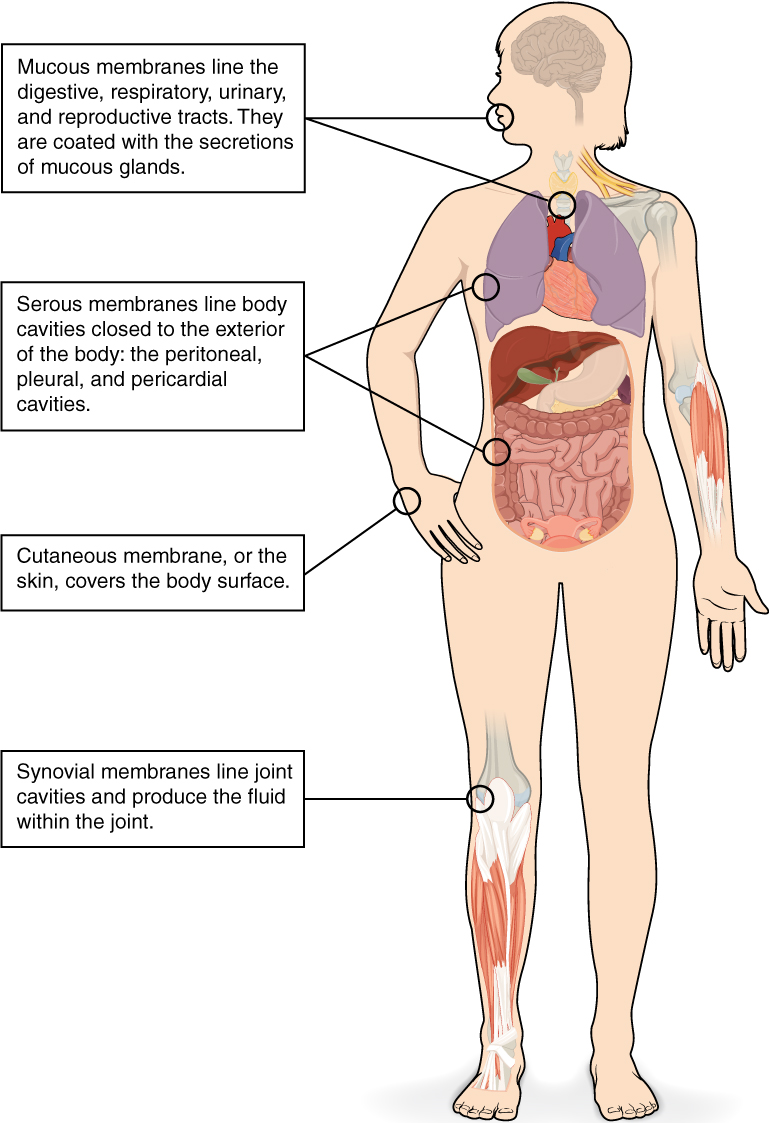
Tissue Membranes
A tissue membrane is a thin layer or sheet of cells that covers the outside of the body (for example, skin), the organs (for example, pericardium), internal passageways that lead to the exterior of the body (for example, abdominal mesenteries), and the lining of the movable joint cavities. There are two basic types of tissue membranes: connective tissue and epithelial membranes (Figure 3.6).
Connective Tissue Membranes
- The connective tissue membrane is formed solely from connective tissue.
- These membranes encapsulate organs, such as the kidneys, and line our movable joints.
- A synovial membrane is a type of connective tissue membrane that lines the cavity of a freely movable joint.
- For example, synovial membranes surround the joints of the shoulder, elbow, and knee.
Epithelial Membranes
- An epithelial membrane is composed of epithelium attached to a layer of connective tissue.
- A mucous membrane is also a composite of connective and epithelial tissues.
- Sometimes called mucosae, these epithelial membranes line the body cavities and hollow passageways that open to the external environment and include the digestive, respiratory, excretory, and reproductive tracts.
- Mucus, produced by the epithelial exocrine glands, covers the epithelial layer.
- The underlying connective tissue, called the lamina propria (literally “own layer”), helps support the fragile epithelial layer.
- The skin is an epithelial membrane also called the cutaneous membrane.
- It is a stratified squamous epithelial membrane resting on top of connective tissue. The apical surface of this membrane is exposed to the external environment and is covered with dead, keratinized cells that help protect the body from desiccation and pathogens.
Membranes of the Anterior (Ventral) Body Cavity
A serous membrane (also referred to as serosa) is an epithelial membrane composed of mesodermally derived epithelium called the mesothelium that is supported by connective tissue.
These membranes line the coelomic cavities of the body, and they cover the organs located within those cavities. They are essentially membranous bags, with mesothelium lining the inside and connective tissue on the outside.
- Parietal layers: line the walls of the body cavity.
- Visceral layer: covers the organs (the viscera).
Between the parietal and visceral layers is a very thin, fluid-filled serous space.
There are three serous cavities and their associated membranes. Serous membranes provide additional protection to the viscera they enclose by reducing friction that could lead to inflammation of the organs.
- Pleura: surrounds the lungs in the pleural cavity and reduces friction between the lungs and the body wall.
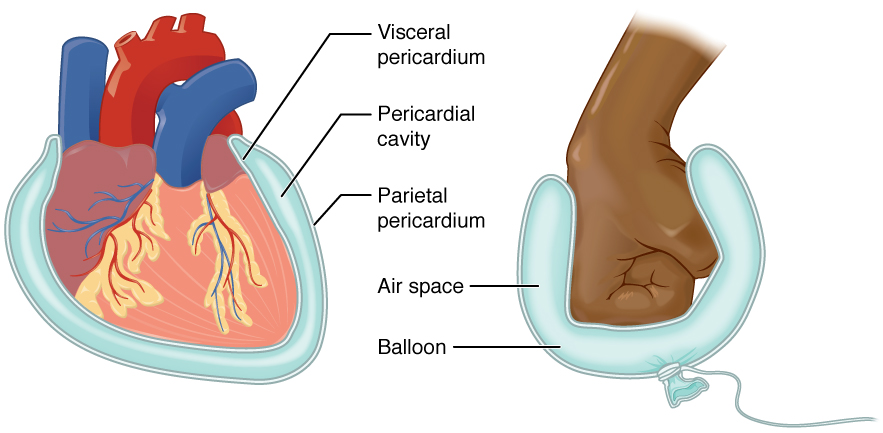
- Pericardium: surrounds the heart in the pericardial cavity and reduces friction between the heart and the wall of the pericardium.
- Peritoneum: surrounds several organs in the abdominopelvic cavity. The peritoneal cavity reduces friction between the abdominal and pelvic organs and the body wall.
***Test yourself
Image Descriptions
Figure 3.5 image description: This illustration shows a lateral and anterior view of the body and highlights the body cavities with different colors. The cranial cavity is a large, bean-shaped cavity filling most of the upper skull where the brain is located. The vertebral cavity is a very narrow, thread-like cavity running from the cranial cavity down the entire length of the spinal cord. Together the cranial cavity and vertebral cavity can be referred to as the dorsal body cavity. The thoracic cavity consists of three cavities that fill the interior area of the chest. The two pleural cavities are situated on both sides of the body, anterior to the spine and lateral to the breastbone. The superior mediastinum is a wedge-shaped cavity located between the superior regions of the two thoracic cavities. The pericardial cavity within the mediastinum is located at the center of the chest below the superior mediastinum. The pericardial cavity roughly outlines the shape of the heart. The diaphragm divides the thoracic and the abdominal cavities. The abdominal cavity occupies the entire lower half of the trunk, anterior to the spine. Just under the abdominal cavity, anterior to the buttocks, is the pelvic cavity. The pelvic cavity is funnel shaped and is located inferior and anterior to the abdominal cavity. Together the abdominal and pelvic cavity can be referred to as the abdominopelvic cavity while the thoracic, abdominal, and pelvic cavities together can be referred to as the ventral body cavity. [Return to Figure 3.5].
Figure 3.6 image description: This illustration shows the silhouette of a human female from an anterior view. Several organs are showing in her neck, thorax, abdomen, left arm, and right leg. Text boxes point out and describe the mucous membranes in several different organs. The topmost box points to the mouth and trachea. It states that mucous membranes line the digestive, respiratory, urinary, and reproductive tracts. They are coated with the secretions of mucous glands. The second box points to the outside edge of the lungs as well as the large intestine and states that serous membranes line body cavities that are closed to the exterior of the body, including the peritoneal, pleural, and pericardial cavities. The third box points to the skin of the hand. It states that the cutaneous membrane, also known as the skin, covers the body surface. The fourth box points to the right knee. It states that synovial membranes line joint cavities and produce the fluid within the joint. [Return to Figure 3.6].
Figure 3.7 image description: This diagram shows the pericardium on the left next to an analogy of a hand punching a balloon on the right. The pericardium is a two-layered sac that surrounds the entire heart except where the blood vessels emerge on the heart’s superior side. The pericardium has two layers because it folds over itself in the shape of the letter U. The inner layer that borders the heart is the visceral pericardium while the outer layer is the parietal pericardium. The space between the two layers is called the pericardial cavity. The heart sits in the cavity much like a fist punching into a balloon. The balloon surrounds the lower part of the fist with a two-layered sac, with the top of the balloon, where it contacts the fist, being analogous to the visceral pericardium. The bottom of the balloon, where it is tied off, is analogous to the parietal pericardium. The air within the balloon is analogous to the pericardial cavity. [Return to Figure 3.7].
