Anatomy and Physiology of the Lymphatic System
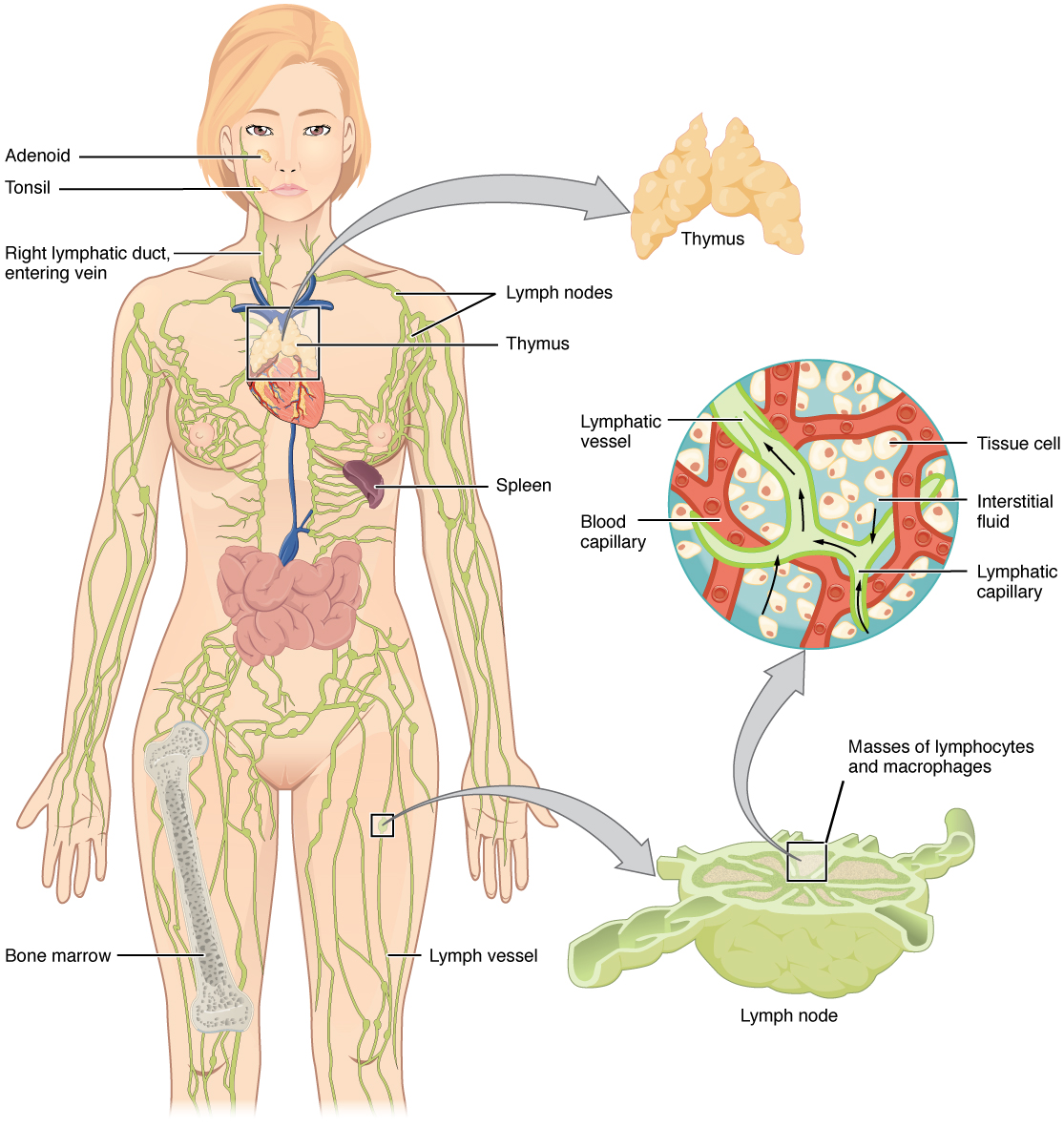
The lymphatic vessels begin as close-ended lymphatic capillaries, which feed into larger and larger lymphatic vessels, and eventually empty into the bloodstream. Along the way, the lymph travels through the lymph nodes, which are commonly found near the groin, armpits, neck, chest, and abdomen. Humans have about 500–600 lymph nodes throughout the body (see Figure 10.1). Several organs and tissues that participate in immunity are also part of the lymphatic system.
Lymphatic Capillaries
An important function of the lymphatic system is to return the fluid (lymph) to the blood. Lymph may be thought of as recycled blood plasma. Blood pressure causes leakage of fluid from the blood capillaries, resulting in the accumulation of fluid in the interstitial space. In humans, 20 liters of plasma are released into the interstitial space of the tissues each day due to capillary leakage. The blood vessels reabsorb 17 liters of this interstitial fluid, leaving 3 liters in the tissues for the lymphatic system to transport back into the body’s circulation. If the lymphatic system is damaged in some way, such as by being blocked by cancer cells or destroyed by injury, interstitial fluid accumulates in the tissue spaces, causing a condition called lymphedema.
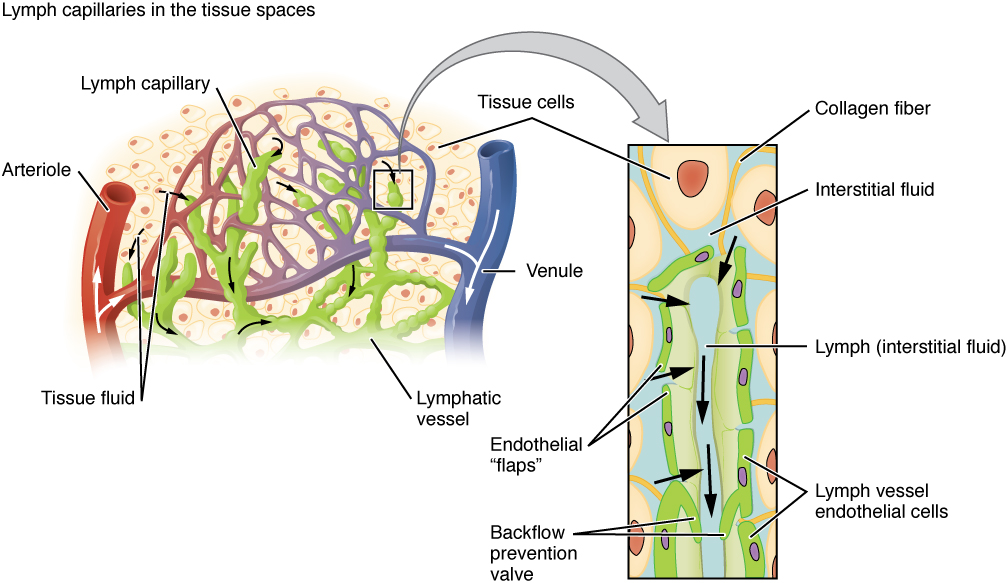
Lymphatic capillaries, also called terminal lymphatics, are vessels where interstitial fluid enters the lymphatic system to become lymph. Located in almost every tissue in the body, these vessels are interlaced among the arterioles and venules of the circulatory system in the soft connective tissues of the body. See Figure 10.2. Exceptions are the central nervous system, bone marrow, bones, teeth, and the cornea of the eye, which do not contain lymph vessels.
Larger Lymphatic Vessels, Trunks, and Ducts
The lymphatic capillaries empty into larger lymphatic vessels, which are similar to veins in terms of their three-tunic structure and the presence of valves. These one-way valves are located fairly close to one another, and each one causes a bulge in the lymphatic vessel (see Figure 10.2).
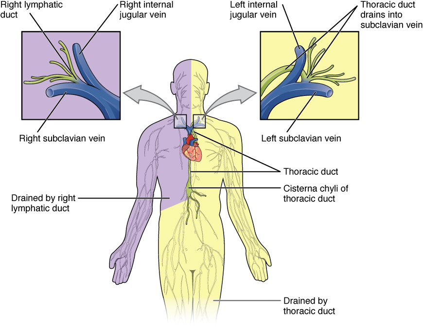
In general, superficial lymphatics follow the same routes as veins, whereas deep lymphatic vessels of the viscera generally follow the paths of arteries. The superficial and deep lymphatics eventually merge to form larger lymphatic structures known as lymphatic trunks. On the right side of the body, the right sides of the head, thorax, and right upper limb trunks drain lymph fluid into the right subclavian vein via the right lymphatic duct (see Figure 10.3). On the left side of the body, the trunks from the remaining portions of the body drain into the larger thoracic duct, which drains into the left subclavian vein. The thoracic duct itself begins just beneath the diaphragm in the cisterna chyli.
Primary Lymphoid Organs
The primary lymphoid organs are the bone marrow and thymus gland. The lymphoid organs are where lymphocytes mature, proliferate, and are selected, which enables them to attack pathogens without harming the cells of the body.
Bone Marrow
Recall that all blood cells, including lymphocytes, are formed in the red bone marrow. The B cell undergoes nearly all of its development in the red bone marrow, whereas the immature T cell, called a thymocyte, leaves the bone marrow and matures largely in the thymus gland.
Thymus
The thymus gland, where T cells mature, is a bilobed organ found in the space between the sternum and the aorta of the heart. Connective tissue holds the lobes closely together but also separates them and forms a capsule.
Secondary Lymphoid Organs
Lymphocytes develop and mature in the primary lymphoid organs, but they mount immune responses from the secondary lymphoid organs, which include the lymph nodes, spleen, and lymphoid nodules. A naïve lymphocyte is one that has left the primary organ, where it learned to function immunologically, and entered a secondary lymphoid organ, where it waits to encounter an antigen against which it will mount a response.
Lymph Nodes
Lymph nodes function to remove debris and pathogens from the lymph, and are thus sometimes referred to as the “filters of the lymph.” Any bacteria that infect the interstitial fluid are taken up by the lymphatic capillaries and transported to a regional lymph node. Dendritic cells and macrophages within this organ internalize and kill many of the pathogens that pass through, thereby removing them from the body. The lymph node is also the site of adaptive immune responses mediated by T cells, B cells, and accessory cells of the adaptive immune system.
Spleen
The spleen is a vascular organ that is somewhat fragile due to the absence of a capsule. It is about 12 cm long and is attached to the lateral border of the stomach. The spleen is sometimes called the “filter of the blood” because of its extensive vascularization and the presence of macrophages and dendritic cells that remove microbes and other materials from the blood, including dying red blood cells. The spleen also functions as the location of immune responses to blood-borne pathogens.
Lymphoid Nodules
The other lymphoid tissues, the lymphoid nodules, consist of a dense cluster of lymphocytes without a surrounding fibrous capsule. These nodules are located in the respiratory and digestive tracts, areas routinely exposed to environmental pathogens. These include tonsils, bronchus-associated lymphoid tissue (BALT), and mucosa-associated lymphoid tissue (MALT).
Image Descriptions
Figure 10.1 image description: The left panel shows a female human body, and the entire lymphatic system is shown Labels read (clockwise from top): thymus, lymph nodes, thymus, spleen, lymph vessel, bone marrow, right lymphatic duct, entering vein, tonsil, adenoid. The right panel shows magnified images of the thymus and the lymph node. Labels read (clockwise from top): tissue cell, interstitial fluid, lymphatic capillary, blood capillary, lymphatic vessel. Label of lymph node reads masses of lymphocytes and macrophages. [Return to Figure 10.1].
Figure 10.2 image description: This image shows the lymph capillaries in the tissue spaces. Labels read (clockwise, from top): lymph capillary, tissue cells, venule, lymphatic vessel, tissue fluid, arteriole. It also shows a magnified image of the interstitial fluid and the lymph vessels. Labels read (clockwise, from top): collagen fiber, interstitial fluid, lymph, lymph vessel endothelial cells, backflow prevention valve, endothelial flaps. [Return to Figure 10.2].
Figure 10.3 image description: This figure shows the lymphatic trunks and the duct system in the human body. Labels read (clockwise from top) thoracic duct, cisterna chyli of thoracic duct, drained by thoracic duct, drained by right lymphatic duct. Callouts to the left and right show the magnified views of the left and right jugular veins, respectively. Labels read (right lymphatic duct): right internal jugular vein, right subclavian vein, right lymphatic duct; (left jugular vein): left internal jugular vein, thoracic duct drains into subclavian vein, left subclavian vein.[Return to Figure 10.3].
