Physiology of the Heart
In order for the heart to do its job of pumping blood to the lungs and to the body, nutrients and oxygen must be supplied to the cells of the heart. The heart also needs to coordinate its contractions so that all parts are working together to pump blood effectively. To understand how all of this works together to give the heart its ability to pump blood, we will examine three interdependent aspects of heart function:
- Circulation through the heart: Blood is pumped by the heart in order to provide oxygen and nutrients to every cell in the body.
- The heart as an organ (coronary blood supply): The heart is an organ made of cells and tissues that require their own blood supply.
- The heart’s electrical conduction system: The heart is able to independently generate and transmit electrical signals to the myocardium in order to make it contract and pump the blood.
1. Circulation through the Heart: The Heart as a Pump
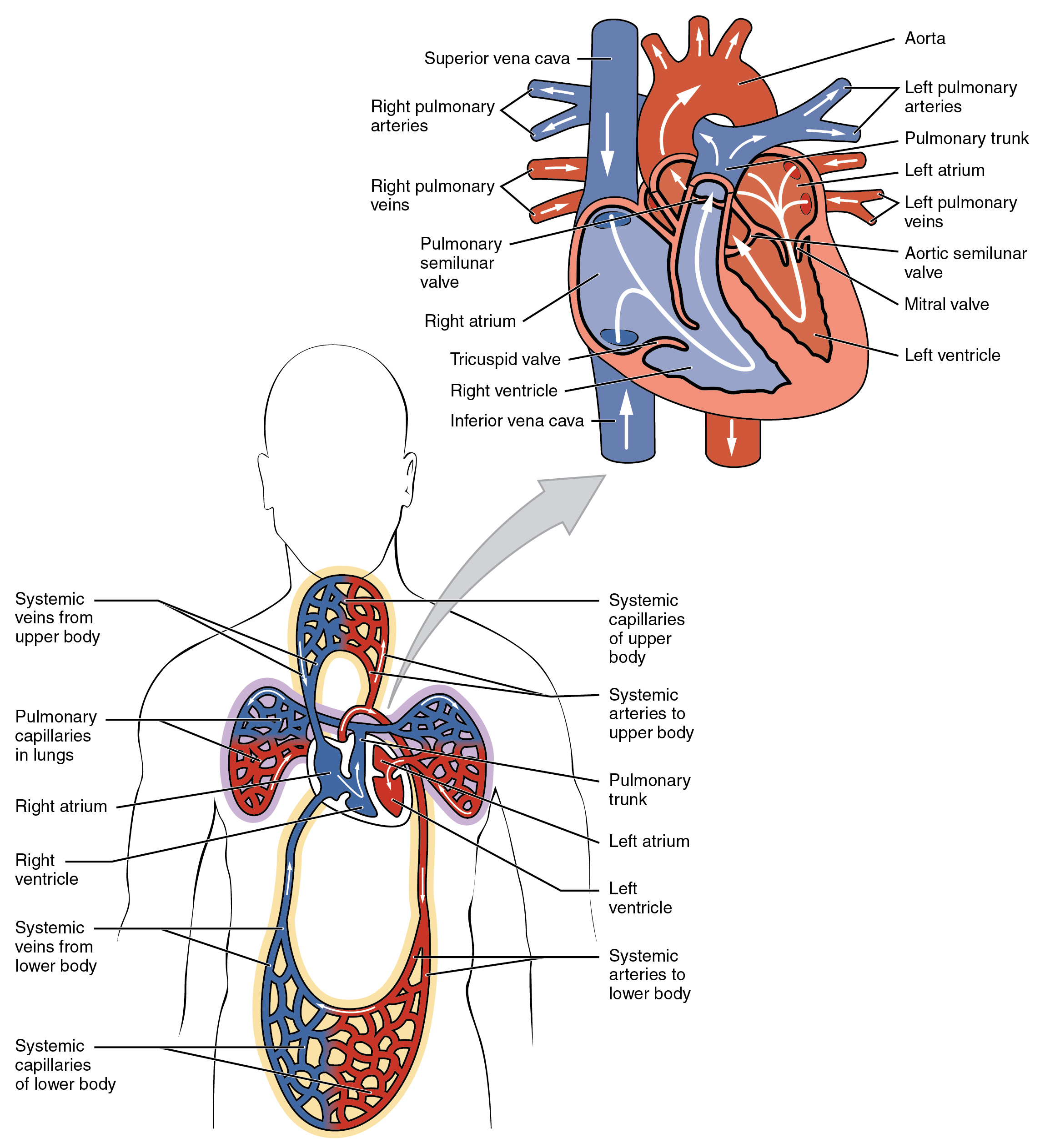
The heart pumps blood to two distinct but linked circulatory systems called the pulmonary and systemic circuits. The pulmonary circuit transports blood to and from the lungs, where it picks up oxygen and drops off carbon dioxide. The systemic circuit transports freshly oxygenated blood to virtually all of the tissues of the body and returns relatively deoxygenated blood and carbon dioxide to the heart to be sent back to the pulmonary circulation.
Cardiac Cycle
The process of pumping and circulating blood is active, coordinated, and rhythmic. Each heartbeat represents one cycle of the heart receiving blood and ejecting blood.
- Diastole is the portion of the cycle in which the heart is relaxed and the atria and ventricles are filling with blood. The AV valves are open so that blood can move from the atria to the ventricles.
- Systole is the portion of the cycle in which the heart contracts, the AV valves slam shut, and the ventricles eject blood to the lungs and to the body through the open semilunar valves. Once this phase ends, the semilunar valves close in preparation for another filling phase.
2. The Heart as an Organ: The Coronary Blood Supply
Myocardial cells require their own blood supply to carry out their function of contracting and relaxing the heart in order to pump blood. This blood supply, provided by the coronary arteries and coronary veins, supplies nutrients and oxygen and carries away carbon dioxide and waste.
3. The Heart’s Electrical Conduction System
In order for all parts of the heart to work together to beat regularly and effectively, the heart has its own electrical system, which initiates and conducts each heartbeat through the entire myocardium. Specialized groups of heart cells perform this function all on their own, without requiring messages from the central nervous system.
Image Descriptions
Figure 8.4 image description: The top panel shows the human heart with the arteries and veins labeled (from top, clockwise): aorta, left pulmonary arteries, pulmonary trunk, left atrium, left pulmonary veins, aortic semilunar valve, mitral valve, left ventricle, inferior vena cava, right ventricle, tricuspid valve, right atrium, pulmonary semilunar valve, right pulmonary veins, right pulmonary arteries, superior vena cava. The bottom panel shows a rough map of the human circulatory system. Labels read (from top, clockwise): systemic capillaries of upper body, systemic arteries to upper body, pulmonary trunk, left atrium, left ventricle, systemic arteries to lower body, systemic capillaries of lower body, systemic veins from lower body, right ventricle, right atrium, pulmonary capillaries in lungs, systemic veins from upper body. [Return to Figure 8.4].
