Basic Anatomy & Physiology of the Musculoskeletal System
The Skeletal System
The skeletal system includes all of the bones, cartilages, and ligaments of the body that support and give shape to the body and body structures. The skeleton consists of the bones of the body. For adults, there are 206 bones in the skeleton. Younger individuals have higher numbers of bones because some bones fuse together during childhood and adolescence to form an adult bone. The primary functions of the skeleton are to provide a rigid, internal structure that can support the weight of the body against the force of gravity and to provide a structure upon which muscles can act to produce movements of the body.
In addition to providing for support and movements of the body, the skeleton has protective and storage functions. It protects the internal organs, including the brain, spinal cord, heart, lungs, and pelvic organs. The bones of the skeleton serve as the primary storage site for important minerals such as calcium and phosphate. The bone marrow found within bones stores fat and houses the blood-cell-producing tissue of the body.
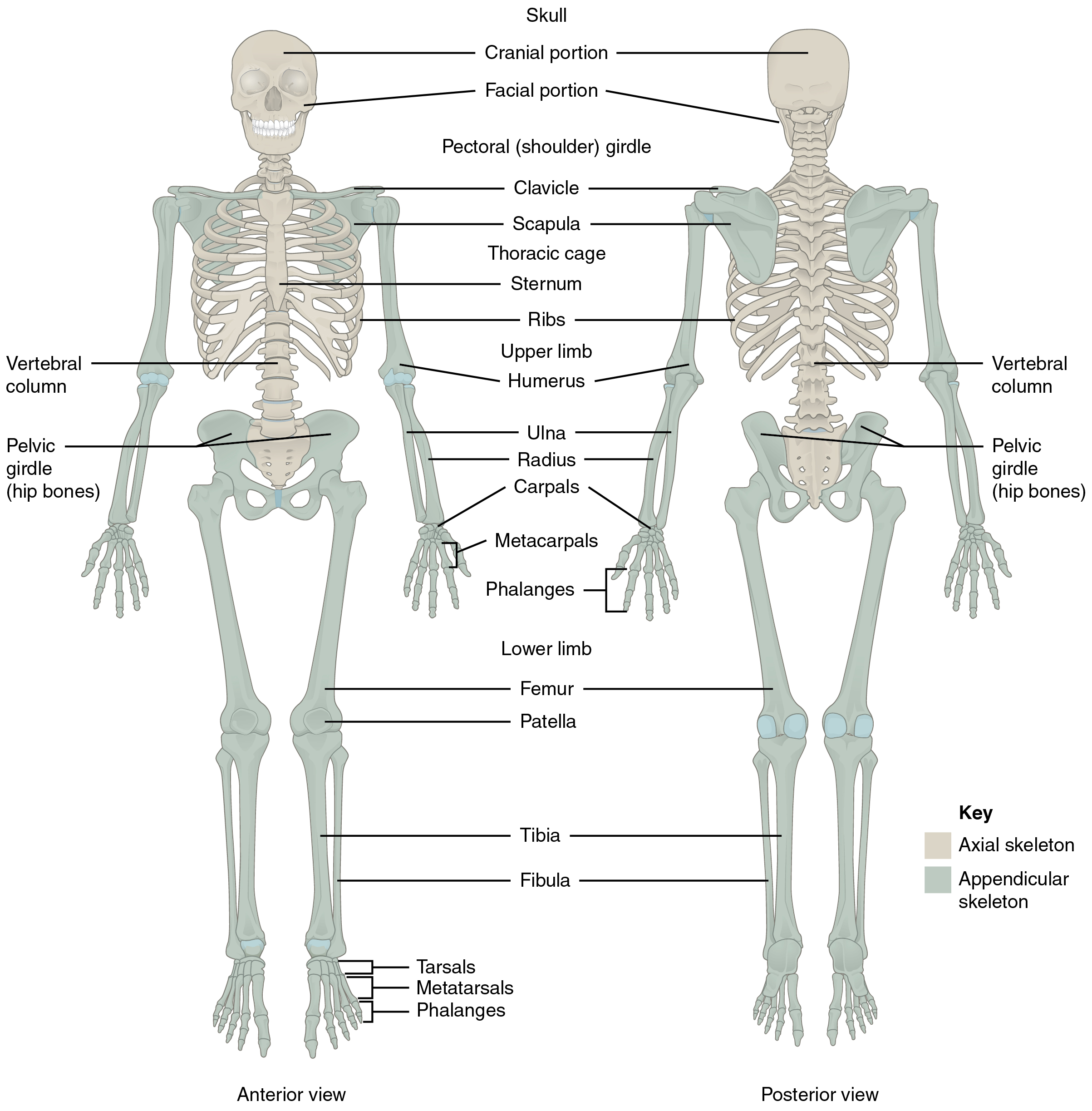
The skeleton is subdivided into two major divisions: the axial and appendicular (see Figure 4.1).
The axial skeleton forms the vertical, central axis of the body and includes all bones of the head, neck, chest, and back. It serves to protect the brain, spinal cord, heart, and lungs. It also serves as the attachment site for muscles that move the head, neck, and back and for muscles that act across the shoulder and hip joints to move their corresponding limbs.
The axial skeleton of the adult consists of 80 bones, including the skull, the vertebral column, and the thoracic cage. The skull is formed by 22 bones. Also associated with the head are an additional seven bones, including the hyoid bone and the ear ossicles (three small bones found in each middle ear). The vertebral column consists of 24 bones, each called a vertebra, plus the sacrum and coccyx. The thoracic cage includes the 12 pairs of ribs and the sternum, the flattened bone of the anterior chest.
Vertebral Column with Abbreviations:
- Cervical: C1 to C7; the first 7 vertebrae in the neck region
- Thoracic: T1 to T12; the next 12 vertebrae that form the outward curvature of the spine
- Lumbar: L1 to L5; the next 5 vertebrae that form the inner curvature of the spine
- Sacrum: the triangular bone at the base of the spine
- Coccyx: the tailbone
The appendicular skeleton includes all bones of the upper and lower limbs, plus the bones that attach each limb to the axial skeleton. There are 126 bones in the appendicular skeleton of an adult.
Most bones connect to at least one other bone in the body. The areas where bones meet bones or where bones meet cartilage are called articulations. Joints can be classified based on their ability to move. At movable joints, the articulating surfaces of the adjacent bones can move smoothly against each other. However, other joints may be connected to each other by connective tissue or cartilage. These joints are designed for stability and provide for little or no movement. Importantly, joint stability and movement are related to each other. This means that stable joints allow for little or no mobility between the adjacent bones. Conversely, joints that provide the most movement between bones are the least stable.
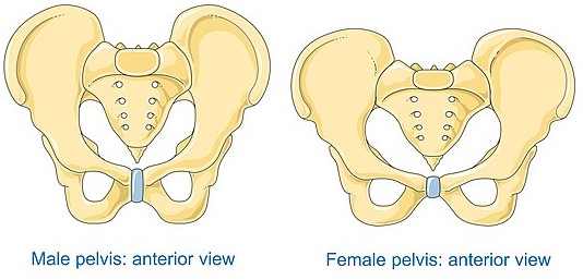
It is important to note the difference between male and female anatomic structures. The pelvis is the only way to differentiate the sex of humans in the skeletal system.
Watch this video:
Media 4.1 Bones, part 1, Crash Course A&P #19 [Online Video]. Copyright 2015 by CrashCourse.
Muscles
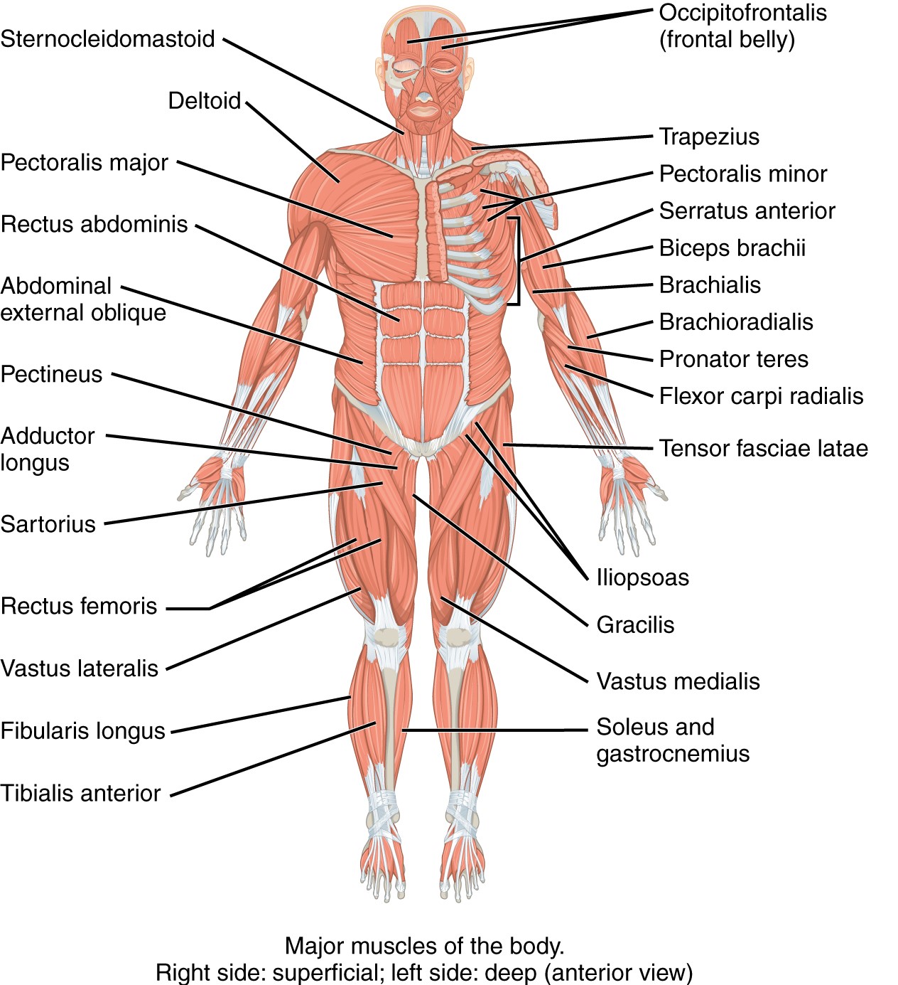
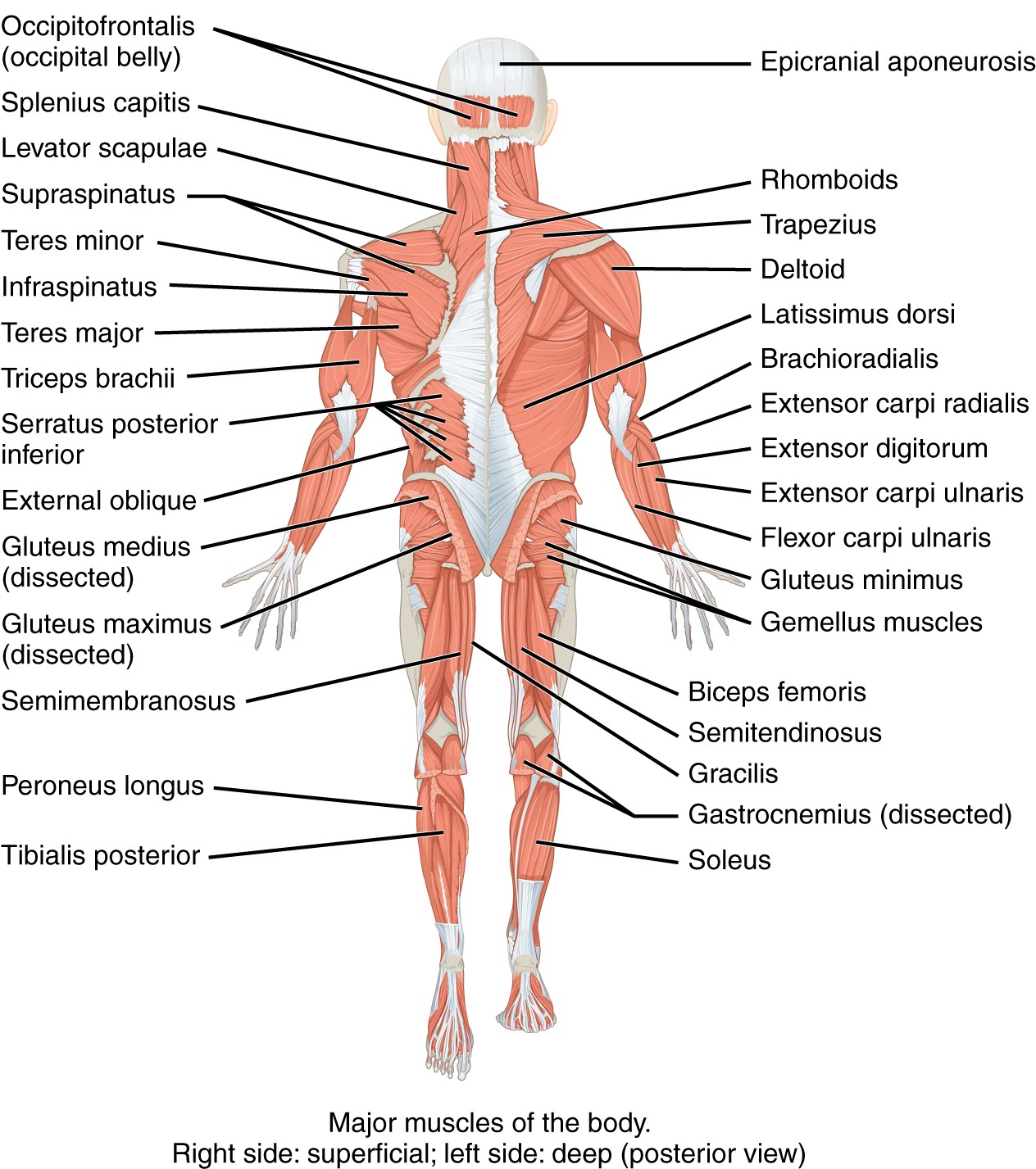
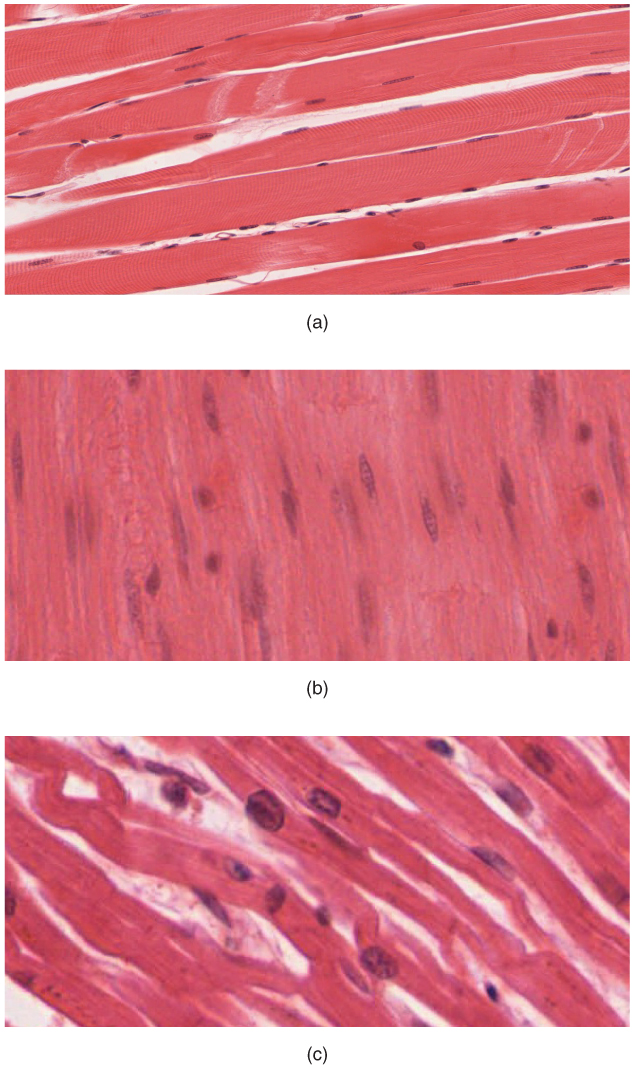
Muscle is one of the four primary tissue types of the body. Muscle tissue covers the entire body (see Figure 4.3 and Figure 4.4) and is made up of specialized cells called fibers.
Types of Muscle Tissue
The body contains three types of muscle tissue: skeletal muscle, cardiac muscle, and smooth muscle (see Figure 4.5). All three muscle tissues have some properties in common; they all exhibit a quality called excitability, as their plasma membranes can change their electrical states (from polarized to depolarized) and send an electrical wave called an action potential along the entire length of the membrane. Fascia is fibrous connective tissue that encloses muscles.
- Skeletal—closely associated with the skeletal system, they are striated muscles and are responsible for voluntary muscle movement—such as swallowing, etc.
- Smooth—mainly associated with the walls of the internal organs. Also known as visceral muscles and are responsible for involuntary muscle movement—such as breathing, etc.
- Cardiac—heart muscle or myocardium. Its striated appearance is similar to a skeletal muscle and is responsible for the pumping of blood. It gives the heartbeat.
Types of Body Movements
Body movement occurs when the bones, joints, and muscles work together (see Figure 4.6 and Figure 4.7). The different types of movement are listed below:
- abduction: moving away from the midline
- adduction: moving toward the midline
- inversion: turning inward
- eversion: turning outward
- extension: movement in which a limb is placed in a straight position, increasing the angle between the bone and the joint
- flexion: movement in which a limb is bent, decreasing the angle between the bone and the joint
- pronation: movement that turns the palm down
- supination: movement that turns the palm up
- rotation: turning around its own axis
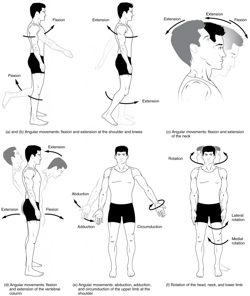
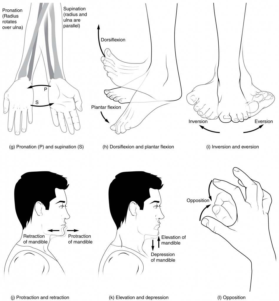
Image Descriptions
Figure 4.1 image description: This diagram shows the human skeleton and identifies the major bones. The left panel shows the anterior view (from the front) and the right panel shows the posterior view (from the back). Labels read (from the top of skull): skull (cranial portion, facial portion), pectoral shoulder girdle, clavicle, scapula, thoracic cage (sternum, ribs), upper limb (humerus, ulna, radius, carpals, metacarpals, phalanges), vertebral column, pelvic girdle (hip bones), lower limb (femur, patella, tibia, fibula, tarsals, metatarsals, phalanges). [Return to Figure 4.1].
Figure 4.3 image description: This diagram shows an anterior view of the muscle system. Labels on the right side of the body read (from top to bottom): sternocleidomastoid, deltoid, pectoralis major, rectus abdominis, abdominal external oblique, pectineus, adductor longus, sartorius, rectus femoris, vastus lateralis, fibularis longus, tibialis anterior. Labels for left side of the body read (from top to bottom): occipitofrontalis (frontal belly), trapezius, pectoralis minor, serratus anterior, biceps brachii, brachialis, brachioradialis, pronator teres, flexor carpi radialis, tensor fasciae latae, iliopsoas, gracilis, vastus medialis, soleus, and gastrocnemius. [Return to Figure 4.3].
Figure 4.4 image description: This diagram shows an anterior view of the muscle system. Labels on the left side of the body read (from top to bottom): occipitofrontalis (occipital belly), splenius capitis, levator scapulae, supraspinatus, teres minor, intraspinatus, teres major, triceps brachii, serratus posterior inferior, external oblique, gluteus medius (dissected), gluteus maximus (dissected) semimembranosus, peroneus longus, tibialis posterior. Labels for right side of the body read (from top to bottom): epicranial aponeurosis, rhomboids, trapezius, deltoid, latissimus dorsi, brachioradialis, extensor carpi radialis, extensor digitorum, extensor carpi ulnaris, flexor carpi ulnaris, gluteus minimus, gemellus muscles, biceps femoris, semitendinosus, gracilis, gastrocnemius (dissected), soleus. [Return to Figure 4.4].
Figure 4.6 image description: This multi-part image shows different types of movements that are possible by different joints in the body. Labels read (from top, left): (a) and (b) angular movements: flexion and extension at the shoulders and knees, (c) angular movements: flexion and extension of the neck (arrows pointing left and right to indicate movement). Labels (from bottom, left) read: (d) angular movements: flexion and extension of the vertical column, (e) angular movements: abduction, adduction, and circumduction of the upper limb at the shoulder, (f) rotation of the head, neck, and lower limb. [Return to Figure 4.6].
