Basic Anatomy & Physiology of the Endocrine System
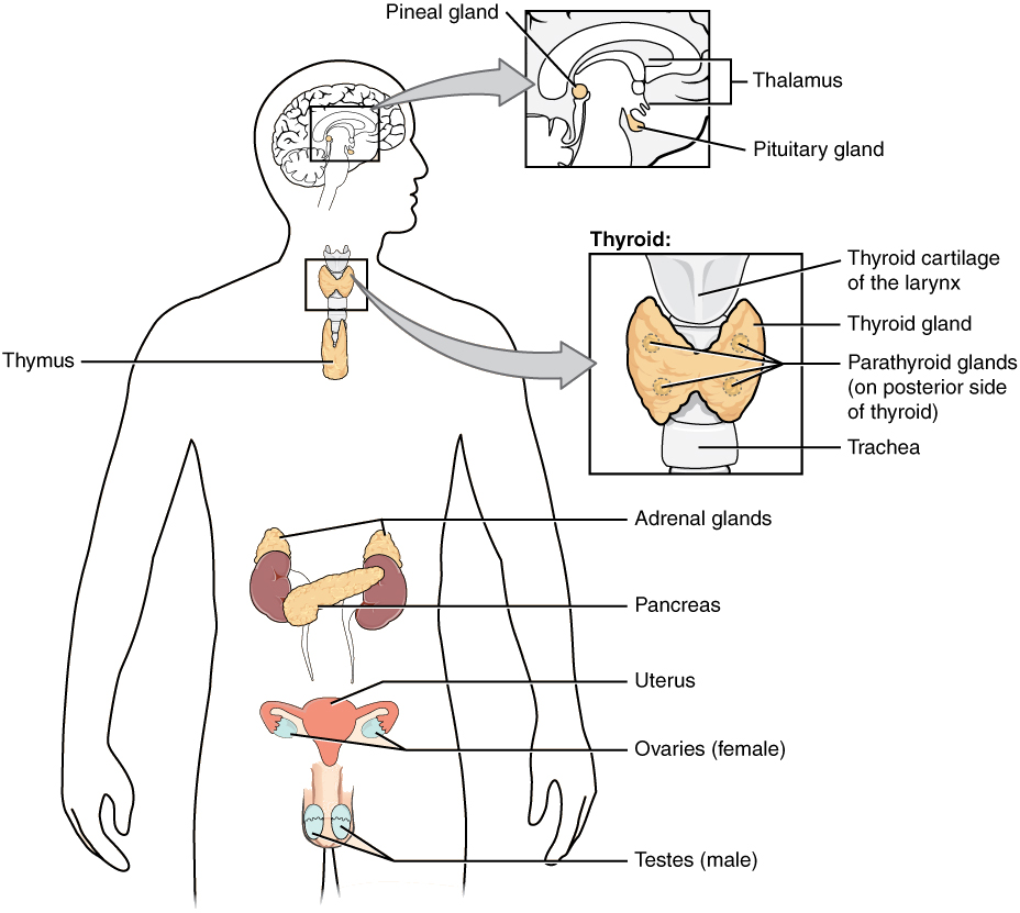
The endocrine system is composed of endocrine glands distributed throughout the body. These glands are pituitary, thyroid, parathyroid, adrenal, pineal, pancreas, gonads (ovaries and testes), and thymus (Figure 6.1).
The endocrine system consists of cells, tissues, and organs that secrete hormones as a primary or secondary function. The primary function of the endocrine glands is to secrete hormones into the bloodstream. Hormones are chemical messengers that will influence metabolic activities, growth, and development. Some glands have both endocrine and nonendocrine functions. For example, the pancreas contains cells that function in digestion as well as cells that secrete the endocrine hormones like insulin and glucagon, which regulate blood glucose levels. The hypothalamus, thymus, heart, kidneys, stomach, small intestine, liver, skin, female ovaries, and male testes are other organs that contain cells with endocrine function. Moreover, fat (adipose) tissue has long been known to produce hormones, and recent research has revealed that even bone tissue has endocrine functions. The ductless endocrine glands are not to be confused with the body’s exocrine system, whose glands release their secretions through ducts. Examples of exocrine glands include the sebaceous and sweat glands of the skin. As just noted, the pancreas also has an exocrine function: most of its cells secrete pancreatic juice through the pancreatic and accessory ducts to the lumen of the small intestine.
Endocrine Signaling
The endocrine system uses one method of communication called chemical signaling. These chemical signals are sent by the endocrine organs. The endocrine organs secrete chemicals—called hormones—into the fluid outside of the tissue cells (extracellular fluid). Hormones are then transported primarily via the bloodstream throughout the body, where they bind to receptors on target cells, creating a particular response. For example, the hormones released when you are presented with a dangerous or a frightening situation, called the fight-or-flight response, occur through the release of hormones from the adrenal gland—epinephrine and norepinephrine—within seconds. In contrast, it may take up to 48 hours for target cells to respond to certain reproductive hormones.
In addition, endocrine signaling is typically less specific than neural (nerve) signaling. The same hormone may also play a role in a variety of different physiological processes depending on the target cells involved. For example, the hormone oxytocin generates uterine contractions in women who are in labor. This hormone is also important in generating the milk-release reflex during breastfeeding and may be involved in the sexual response and in feelings of emotional attachment in both males and females.
Generally, the nervous system involves quick responses to rapid changes in the external environment, and the endocrine system is usually slower acting—taking care of the internal environment of the body, maintaining equilibrium (homeostasis), and controlling reproduction (see Table 6.1). So how does the fight-or-flight response, mentioned earlier, happen so quickly if hormones are usually slower acting? It is because the two systems are connected. It is the fast action of the nervous system in response to the danger in the environment that stimulates the adrenal glands to secrete their hormones, epinephrine and norepinephrine. As a result, the nervous system can cause rapid endocrine responses to keep up with sudden changes in both the external and internal environments when necessary.
Table 6.1: Endocrine and Nervous Systems
| Characteristic | Endocrine System | Nervous System |
|---|---|---|
| Signaling mechanism(s) | Chemical | Chemical/electrical |
| Primary chemical signal | Hormones | Neurotransmitters |
| Distance traveled | Long or short | Always short |
| Response time | Fast or slow | Always fast |
| Environment targeted | Internal | Internal and external |
From Betts, et al., 2013. Licensed under CC BY 4.0.
Table 6.2: Endocrine Glands and Their Major Hormones
| Endocrine Gland | Associated Hormones | Chemical Class | Effect |
|---|---|---|---|
| Pituitary (anterior) | Growth hormone (GH) | Protein | Promotes growth of body tissues |
| Pituitary (anterior) | Prolactin (PRL) | Peptide | Promotes milk production |
| Pituitary (anterior) | Thyroid-stimulating hormone (TSH) | Glycoprotein | Stimulates thyroid hormone release |
| Pituitary (anterior) | Adrenocorticotropic hormone (ACTH) | Peptide | Stimulates hormone release by adrenal cortex |
| Pituitary (anterior) | Follicle-stimulating hormone (FSH) | Glycoprotein | Stimulates gamete production |
| Pituitary (anterior) | Luteinizing hormone (LH) | Glycoprotein | Stimulates androgen production by gonads |
| Pituitary (posterior) | Antidiuretic hormone (ADH) | Peptide | Stimulates water reabsorption by kidneys |
| Pituitary (posterior) | Oxytocin | Peptide | Stimulates uterine contractions during childbirth |
| Thyroid | Thyroxine (T4), triiodothyronine (T3) | Amine | Stimulate basal metabolic rate |
| Thyroid | Calcitonin | Peptide | Reduces blood Ca2+ levels |
| Parathyroid | Parathyroid hormone (PTH) | Peptide | Increases blood Ca2+ levels |
| Adrenal (cortex) | Aldosterone | Steroid | Increases blood Na+ levels |
| Adrenal (cortex) | Cortisol, corticosterone, cortisone | Steroid | Increases blood glucose levels |
| Adrenal (medulla) | Epinephrine, norepinephrine | Amine | Stimulates fight-or-flight response |
| Pineal | Melatonin | Amine | Regulates sleep cycles |
| Pancreas | Insulin | Protein | Reduces blood glucose levels |
| Pancreas | Glucagon | Protein | Increases blood glucose levels |
| Testes | Testosterone | Steroid | Stimulates development of male secondary sex characteristics and sperm production |
| Ovaries | Estrogens and progesterone | Steroid | Stimulates development of female secondary sex characteristics and prepares the body for childbirth |
From Betts, et al., 2013. Licensed under CC BY 4.0.
Hormone Receptors
The message a hormone sends is received by a hormone receptor, a protein located either inside the cell or within the cell membrane. The receptor will process the message by initiating other signaling events or cellular mechanisms that result in the target cell’s response. Hormone receptors recognize molecules with specific shapes and side groups and respond only to those hormones that are recognized. The same type of receptor may be located on cells in different body tissues and trigger somewhat different responses. Thus, the response triggered by a hormone depends not only on the hormone but also on the target cell.
Image Descriptions
Figure 6.1 image description: This diagram shows the endocrine glands and cells that are located throughout the body. The endocrine system organs include the pineal gland and pituitary gland in the brain. The pituitary is located on the anterior side of the thalamus, while the pineal gland is located on the posterior side of the thalamus. The thyroid gland is a butterfly-shaped gland that wraps around the trachea within the neck. Four small, disc-shaped parathyroid glands are embedded into the posterior side of the thyroid. The adrenal glands are located on top of the kidneys. The pancreas is located at the center of the abdomen. In females, the two ovaries are connected to the uterus by two long, curved tubes in the pelvic region. In males, the two testes are located in the scrotum below the penis. [Return to Figure 6.1].
