Anatomy of the Nervous System
The Central and Peripheral Nervous Systems
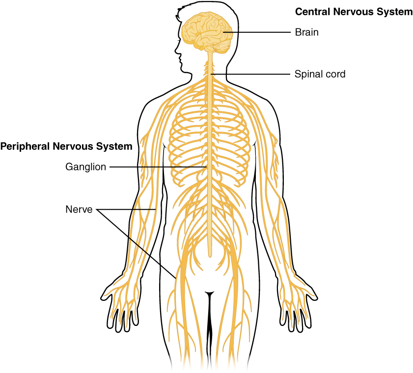
The nervous system can be divided into two major regions: the central and peripheral nervous systems. The central nervous system (CNS) is the brain and spinal cord, and the peripheral nervous system (PNS) is everything else (see Figure 5.1). The brain is contained within the cranial cavity of the skull, and the spinal cord is contained within the vertebral cavity of the vertebral column. The peripheral nervous system is so named because it is on the periphery—meaning beyond the brain and spinal cord.
Nervous tissue, present in both the CNS and PNS, contains two basic types of cells: neurons and glial cells. Neurons are the primary type of cell that most anyone associates with the nervous system. They are electrically active and release chemical signals to target cells. A glial cell is one of a variety of cells that provide a framework of tissue that supports the neurons and their activities.
Neurons are cells and therefore have a soma, or cell body, but they also have extensions of the cell; each extension is generally referred to as a process. There is one important process that every neuron has called an axon, which is the fiber that connects a neuron with its target. Another type of process that branches off from the soma is the dendrite. Dendrites are responsible for receiving most of the input from other neurons.
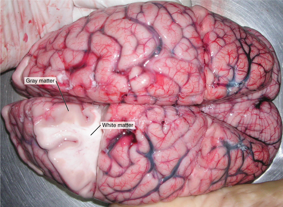
Looking at nervous tissue, there are regions that predominantly contain cell bodies and regions that are largely composed of just axons. These two regions within nervous system structures are often referred to as gray matter (the regions with many cell bodies and dendrites) or white matter (the regions with many axons). Figure 5.2 demonstrates the appearance of these regions in the brain and spinal cord.
The Adult Brain
The adult brain is separated into four major regions: the cerebrum, the diencephalon, the brain stem, and the cerebellum.
The Cerebrum
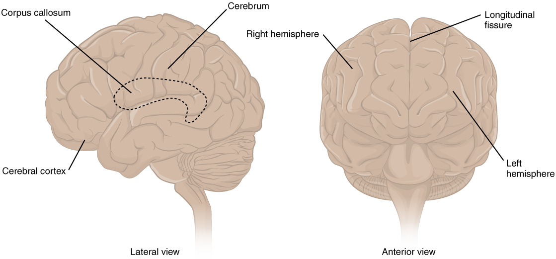
The iconic gray mantle of the human brain, which appears to make up most of the mass of the brain, is the cerebrum (see Figure 5.3). The wrinkled portion is the cerebral cortex, and the rest of the structure is beneath that outer covering. There is a large separation between the two sides of the cerebrum called the longitudinal fissure. It separates the cerebrum into two distinct halves, a right and left cerebral hemisphere. Deep within the cerebrum, the white matter of the corpus callosum provides the major pathway for communication between the two hemispheres of the cerebral cortex.
Many of the higher neurological functions, such as memory, emotion, and consciousness, are the result of cerebral function. In mammals, the cerebrum comprises the outer gray matter that is the cortex (from the Latin word meaning “bark of a tree”).
Cerebral Cortex
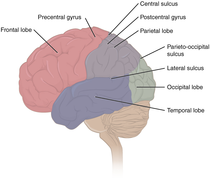
The cerebrum is covered by a continuous layer of gray matter that wraps around either side of the forebrain—the cerebral cortex. This thin, extensive region of wrinkled gray matter is responsible for the higher functions of the nervous system. A gyrus (plural = gyri) is the ridge of one of those wrinkles, and a sulcus (plural = sulci) is the groove between two gyri. The pattern of these folds of tissue indicates specific regions of the cerebral cortex.
Extensive folding in the cerebral cortex enables more gray matter to fit into this limited space. If the gray matter of the cortex were peeled off of the cerebrum and laid out flat, its surface area would be roughly equal to one square meter.
Using these landmarks (the largest gyri and sulci), the cortex can be separated into four major regions, or lobes (see Figure 5.4).
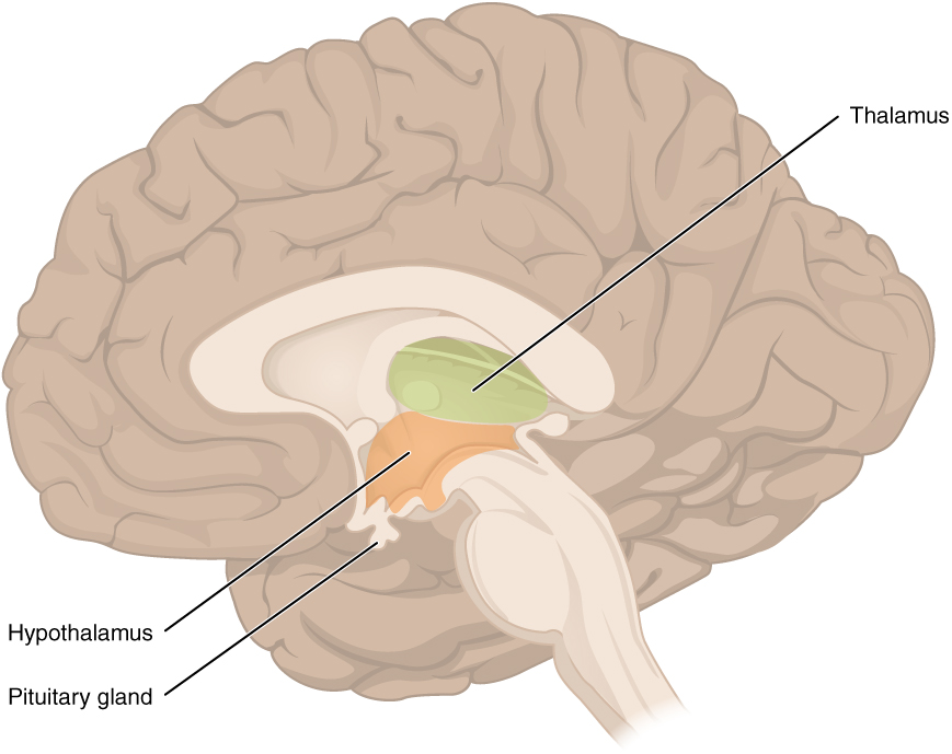
The Diencephalon
The diencephalon is deep beneath the cerebrum and constitutes the walls of the third ventricle. The two major regions of the diencephalon are the thalamus itself and the hypothalamus (see Figure 5.5).
Thalamus
The thalamus is a collection of nuclei that relay information between the cerebral cortex and the periphery, spinal cord, or brain stem. All sensory information, except for the sense of smell, passes through the thalamus before being processed by the cortex.
Hypothalamus
Inferior and slightly anterior to the thalamus is the hypothalamus, the other major region of the diencephalon. The hypothalamus is a collection of nuclei that are largely involved in regulating homeostasis. The hypothalamus is the executive region in charge of the autonomic nervous system and the endocrine system through its regulation of the anterior pituitary gland. Other parts of the hypothalamus are involved in memory and emotion as part of the limbic system.
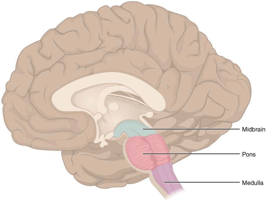
The midbrain and hindbrain (composed of the pons and the medulla) are collectively referred to as the brain stem (see Figure 5.6). The structure emerges from the ventral surface of the forebrain as a tapering cone that connects the brain to the spinal cord. The midbrain coordinates sensory representations of the visual, auditory, and somatosensory perceptual spaces. The pons is the main connection with the cerebellum. The pons and the medulla regulate several crucial functions, including the cardiovascular and respiratory systems and rates. The major ascending and descending pathways between the spinal cord and brain, specifically the cerebrum, pass through the brain stem.
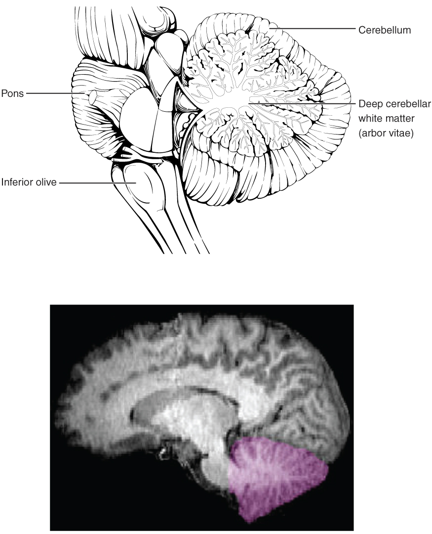
The Cerebellum
The cerebellum, as the name suggests, is the “little brain.” It is covered in gyri and sulci like the cerebrum and looks like a miniature version of that part of the brain (see Figure 5.7). The cerebellum is largely responsible for comparing information from the cerebrum with sensory feedback from the periphery through the spinal cord. It accounts for approximately 10 percent of the mass of the brain.
The Spinal Cord
The description of the CNS is concentrated on the structures of the brain, but the spinal cord is another major organ of the system.
The length of the spinal cord is divided into regions that correspond to the regions of the vertebral column. The name of a spinal cord region corresponds to the level at which spinal nerves pass through the intervertebral foramina. Immediately adjacent to the brain stem are the following divisions of the spinal cord:
- cervical region
- thoracic region
- lumbar region
- sacral region
The spinal cord is not the full length of the vertebral column because the spinal cord does not grow significantly longer after the first or second year, but the skeleton continues to grow. The nerves that emerge from the spinal cord pass through the intervertebral foramina at the respective levels. As the vertebral column grows, these nerves grow with it and result in a long bundle of nerves that resembles a horse’s tail and is named the cauda equina. The sacral spinal cord is at the level of the upper lumbar vertebral bones. The spinal nerves extend from their various levels to the proper level of the vertebral column.
Neurons
Neurons are the cells considered to be the basis of nervous tissue. They are responsible for the electrical signals that communicate information about sensations and that produce movements in response to those stimuli, along with inducing thought processes within the brain.
Parts of a Neuron
As you learned in the first section, the main part of a neuron is the cell body, which is also known as the soma (soma = “body”). The cell body contains the nucleus and most of the major organelles. But what makes neurons special is that they have many extensions of their cell membranes, which are generally referred to as processes. Neurons are usually described as having one, and only one, axon—a fiber that emerges from the cell body and projects to target cells. That single axon can branch repeatedly to communicate with many target cells. It is the axon that propagates the nerve impulse, which is communicated to one or more cells. The other processes of the neuron are dendrites, which receive information from other neurons at specialized areas of contact called synapses. The dendrites are usually highly branched processes, providing locations for other neurons to communicate with the cell body. Information flows through a neuron from the dendrites, across the cell body, and down the axon.
Figure 5.8 shows the relationship of these parts to one another.
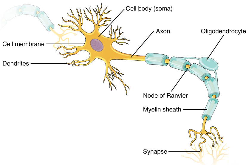
Many axons are wrapped by an insulating substance called myelin, which is actually made from glial cells. Myelin acts as insulation, much like the plastic or rubber that is used to insulate electrical wires. A key difference between myelin and the insulation on a wire is that there are gaps in the myelin covering of an axon. Each gap is called a node of Ranvier and is important to the way that electrical signals travel down the axon. At the end of the axon is the axon terminal, where there are usually several branches extending toward the target cell, each of which ends in an enlargement called a synaptic end bulb. These bulbs are what make the connection with the target cell at the synapse.
Glial Cells
Glial cells, or neuroglia or simply glia, are the other type of cell found in nervous tissue. They are considered to be supporting cells, and many functions are directed at helping neurons complete their function for communication. The name glia comes from the Greek word that means “glue.”
There are six types of glial cells. Four of them are found in the CNS, and two are found in the PNS. Table 5.1 outlines some common characteristics and functions.
Table 5.1: Glial Cell Types by Location and Basic Function
| CNS GLIA | PNS GLIA | BASIC FUNCTION |
|---|---|---|
| Astrocyte | Satellite cell | Support |
| Oligodendrocyte | Schwann cell | Insulation, myelination |
| Microglia | – | Immune surveillance and phagocytosis |
| Ependymal cell | – | Creating CSF |
From Betts, et al., 2013. Licensed under CC BY 4.0.
Image Descriptions
Figure 5.1 image description: This diagram shows a silhouette of a human highlighting the nervous system. The central nervous system is composed of the brain and spinal cord. The brain is a large mass of ridged and striated tissue within the head. The spinal cord extends down from the brain and travels through the torso, ending in the pelvis. Pairs of enlarged nervous tissue, labeled ganglia, flank the spinal cord as it travels through the rib area. The ganglia are part of the peripheral nervous system, along with the many thread-like nerves that radiate from the spinal cord and ganglia through the arms, abdomen, and legs. [Return to Figure 5.1].
Figure 5.2 image description: This photo shows an enlarged view of the dorsal side of a human brain. The right side of the occipital lobe has been shaved to reveal the white and gray matter beneath the surface blood vessels. The white matter branches through the shaved section like the limbs of a tree. The gray matter branches and curves on the outside of the white matter, creating a buffer between the outer edges of the occipital lobe and the internal white matter. [Return to Figure 5.2].
Figure 5.3 image description: This figure shows the lateral view on the left panel and anterior view on the right panel of the brain. The major parts including the cerebrum are labeled. Lateral view labels (clockwise from top) read: cerebrum, cerebral cortex, corpus callosum (located on the interior of the brain). Anterior view labels indicate the right and left hemispheres and the longitudinal fissure between them. [Return to Figure 5.3].
Figure 5.4 image description: This figure shows the lateral view of the brain and the major lobes are labeled. From the front of the brain (left) labels read: frontal lobe, precentral gyrus, central sulcus, postcentral gyrus, parietal lobe, lateral sulcus, occipital lobe, temporal lobe. [Return to Figure 5.4].
Figure 5.5 image description: This figure shows the location of the thalamus, hypothalamus, and pituitary gland in the brain. Each part is labeled respectively. The thalamus is located in the midsection of the brain. The hypothalamus is located below the thalamus and the pituitary gland below that. [Return to Figure 5.5].
Figure 5.6 image description: This figure shows the location of the midbrain, pons, and the medulla in the brain that make up the brain stem. The midbrain is located at the top, the pons is located beneath that, and the medulla is the lowest most point of the brain stem. [Return to Figure 5.6].
Figure 5.7 image description: This figure shows the location of the cerebellum in the brain which is located on the posterior surface of the brain stem. Labels read (top, left): pons, inferior olive, (top, right) cerebellum, deep cerebellar white matter (arbor vitae). In the top panel, a lateral view labels the location of the cerebellum and the deep cerebellar white matter. In the bottom panel, a photograph of a brain, with the cerebellum in pink is shown. [Return to Figure 5.7].
Figure 5.8 image description: This illustration shows the anatomy of a neuron. The neuron has a very irregular cell body (soma) containing a purple nucleus. There are six projections protruding from the top, bottom, and left side of the cell body. Each of the projections branches many times, forming small, tree-shaped structures protruding from the cell body. The right side of the cell body tapers into a long cord called the axon. The axon is insulated by segments of myelin sheath, which resemble a semitransparent toilet paper roll wound around the axon. The myelin sheath is not continuous but is separated into equally spaced segments. The bare axon segments between the sheath segments are called nodes of Ranvier. An oligodendrocyte is reaching its two arm-like projections onto two myelin sheath segments. The axon branches many times at its end, where it connects to the dendrites of another neuron. Each connection between an axon branch and a dendrite is called a synapse. The cell membrane completely surrounds the cell body, dendrites, and its axon. The axon of another nerve is seen in the upper left of the diagram connecting with the dendrites of the central neuron. [Return to Figure 5.8].
